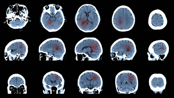
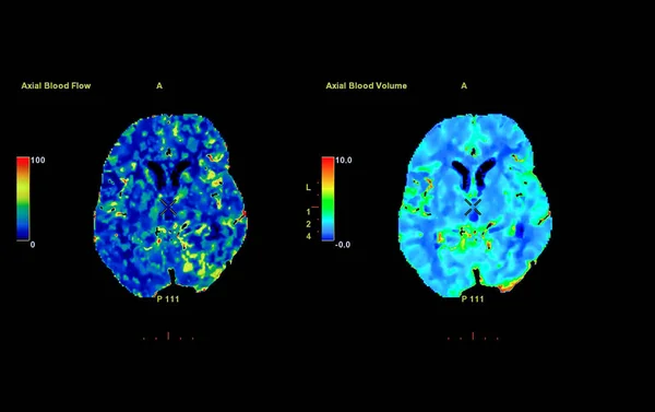
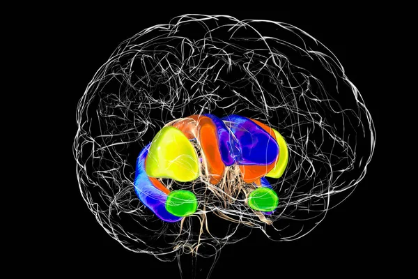
Stock image selective focus of CT Brain Perfusion or CT scan image of the brain 3d rendering image analyzing cerebral blood flow on the monitor.

Published: Dec.23, 2019 11:44:45
Author: samunella
Views: 10
Downloads: 0
File type: image / jpg
File size: 2.15 MB
Orginal size: 5059 x 3436 px
Available sizes:
Level: beginner