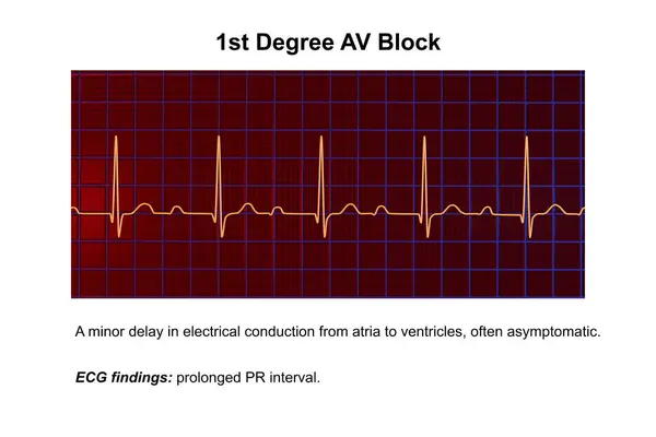
Stock image 3D illustration of an ECG displaying 1st degree AV block, a cardiac conduction disorder.
Published: Mar.04, 2024 11:12:10
Author: katerynakon
Views: 2
Downloads: 1
File type: image / jpg
File size: 10.7 MB
Orginal size: 9000 x 6000 px
Available sizes:
Level: silver