Home
Images
Vectors
Videos
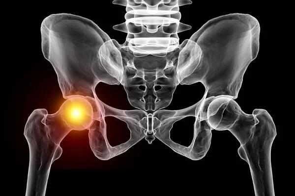
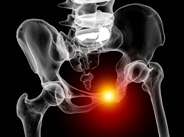
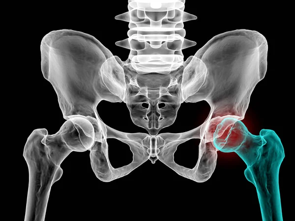
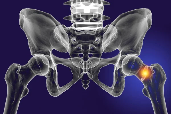
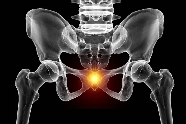
Stock image A 3D medical illustration highlighting the coccyx bone marked in red, depicting coccyx pain which can occur due to injury, childbirth, or prolonged sitting. Front view
Published:
Apr.04, 2023 11:54:01
Author:
katerynakon
Views:
43
Downloads:
2
File type:
image
/
jpg
File size:
4.46 MB
Orginal size:
7000
x
4667
px
Available sizes:
S
(500 x 333)
M
(1000 x 667)
L
(2000 x 1333)
XL
(7000 x 4667)
EL0
(7000 x 4667)
Level:
silver
Download Image
Image topics:
Illustration
Isolated
Sitting
Medical
3d
Back
Structure
Bone
Support
System
Pain
Spine
Joint
Damage
Fracture
Removal
Anatomy
X Ray
Surgery
Trauma
Injury
Alignment
Function
Rehabilitation
Lower
Childbirth
Skeletal
Inflammation
Syndrome
Orthopedics
Nerve
Pelvic
Disorders
Vertebrae
Coccyx
Abscess
Dislocation
Coccygeal
Black Background
Bone Structure
Skeletal System
Lower Back Pain
Tailbone
Coccydynia
Tailbone Pain
Sacrococcygeal
Coccyx Removal
Prolonged Sitting
Coccygeal Pain
Similar stock images
Hip Joint Pain. A 3D Medical Illustration Depicting The Pelvis And Femur Bones With The Femur Head Highlighted In Red, Illustrating Femur Joint Pain Concept.
7000 × 4667
Symphysis Pubis Dysfunction, 3D Illustration Showing Pelvic Bones
6000 × 4494
Femur Bone Affected By Legg-Calve-Perthes Disease, A Childhood Hip Disorder That Affects The Blood Supply To The Femoral Head, 3D Illustration Shows Affected Left Femur Bone (right Side Of The Image)
7000 × 5250
Symphysis Pubis Dysfunction, 3D Illustration Showing Pelvic Bones And Highlighted In Red Pubis Symphysis
6000 × 4494
Female Reproductive System Anatomy. 3D
3840 × 2160
A Fracture Of The Femur Neck, A Common Type Of Hip Fracture That Typically Occurs In Older Adults And Can Lead To Mobility Issues And Other Complications, Isolated On White Background, 3D Illustration
7000 × 4667