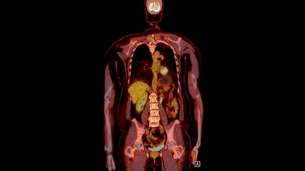
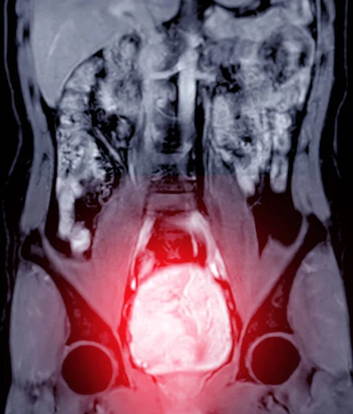
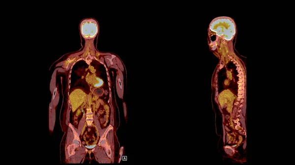
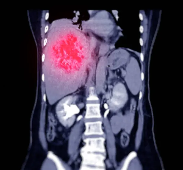
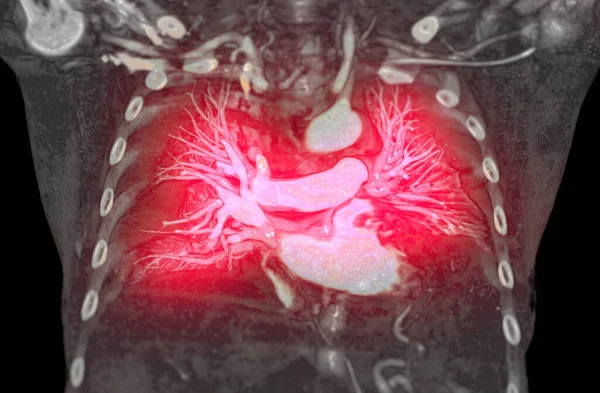
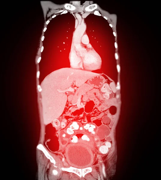
Stock image A PET-CT scan image is a diagnostic visualization combining Positron Emission Tomography (PET) and Computed Tomography (CT) for Helps in finding cancer recurrence.

Published: May.02, 2024 08:48:44
Author: samunella
Views: 0
Downloads: 0
File type: image / jpg
File size: 3.52 MB
Orginal size: 4096 x 5148 px
Available sizes:
Level: beginner