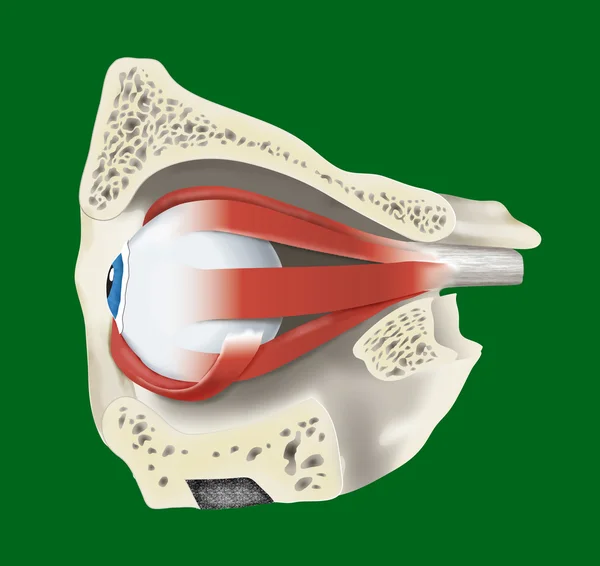
Stock image Anatomic illustration of an eye
Published: May.23, 2012 14:44:42
Author: alexonline
Views: 414
Downloads: 7
File type: image / jpg
File size: 1.6 MB
Orginal size: 4252 x 4016 px
Available sizes:
Level: bronze
Similar stock images
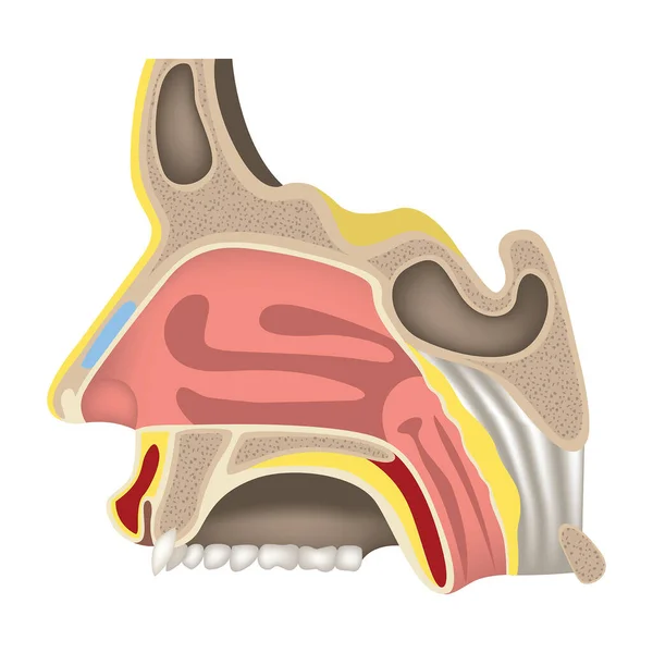
The Nasal Cavity. Charm Organs. Human Head Anatomy. Haimar's Sinus. Profile Cut. Vector Illustration.
2000 × 2000