photo free
Home Images Vectors Videos

Stock image anatomy of carpal tunnel syndrome, highlighting the median nerve, flexor tendons, and carpal bones diagram hand drawn schematic raster illustration. Medical science educational illustration

Stock image anatomy of carpal tunnel syndrome, highlighting the median nerve, flexor tendons, and carpal bones diagram hand drawn schematic raster illustration. Medical science educational illustration
Published: Jul.03, 2024 13:25:26
Author: AlexanderPokusay
Views: 0
Downloads: 0
File type: image / jpg
File size: 4.36 MB
Orginal size: 6000 x 4500 px
Available sizes:
S
(500 x 375)
M
(1000 x 750)
L
(2000 x 1500)
XL
(6000 x 4500)
EL0
(6000 x 4500)
Level: silver
Download Image
Image topics:

Similar stock images

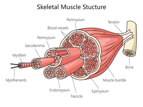
stock image Skeletal anatomy muscle structure medical diagram schematic raster illustration. Medical science educational illustration

Skeletal Anatomy Muscle Structure Medical Diagram Schematic Raster Illustration. Medical Science Educational Illustration

6000 × 4500
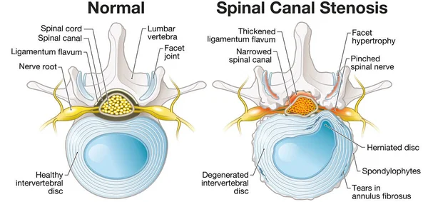
stock image Illustration showing spinal canal stenosis lumbar vertebra with intervertebral disc and herniated nucleus pulposus

Illustration Showing Spinal Canal Stenosis Lumbar Vertebra With Intervertebral Disc And Herniated Nucleus Pulposus

7422 × 3504
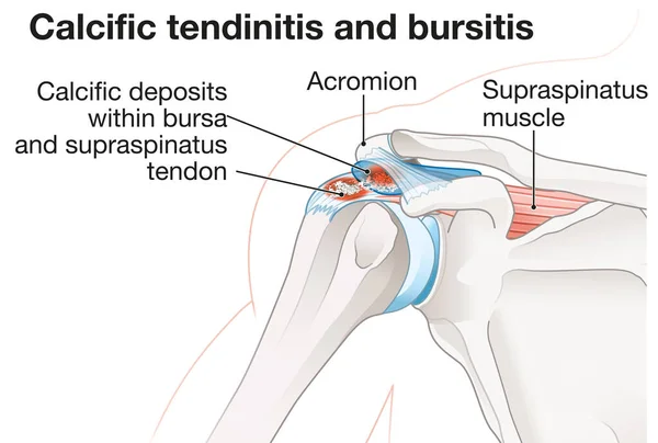
stock image Illustration showing calcific tendinitis and calcific bursitis

Illustration Showing Calcific Tendinitis And Calcific Bursitis

5260 × 3543
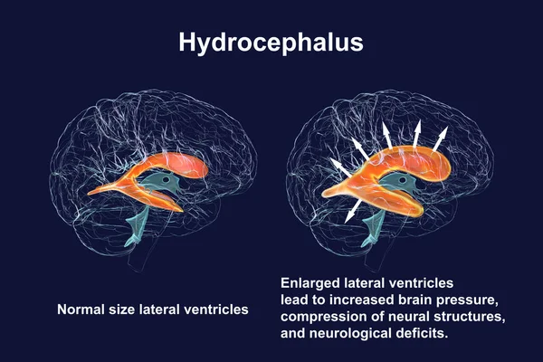
stock image A 3D scientific illustration depicting enlarged lateral ventricles of the child brain (hydrocephalus, right side), and normal ventricular system (left side).

A 3D Scientific Illustration Depicting Enlarged Lateral Ventricles Of The Child Brain (hydrocephalus, Right Side), And Normal Ventricular System (left Side).

7800 × 5200
stock image Spinal Cord Segment. Labeled 3D illustration

Spinal Cord Segment. Labeled 3D Illustration

5506 × 3002