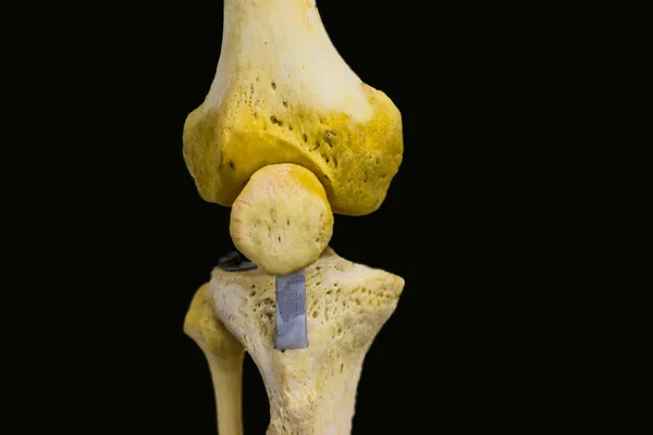
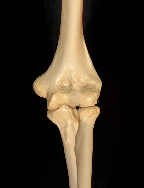
Stock image antero posterior view of articulated femur tibia fibula patella bones showing human knee joint anatomy in isolated black background with space for text
Published: May.15, 2018 19:52:17
Author: shibu7213.gmail.com
Views: 97
Downloads: 6
File type: image / jpg
File size: 3.9 MB
Orginal size: 5184 x 3456 px
Available sizes:
Level: bronze
Similar stock images
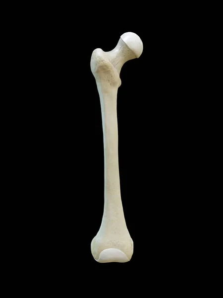
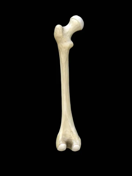
Left Human Femur Bone, Posterior View, Close Up Isolated On Black Background, 3d Rendering
3375 × 4500