Home
Images
Vectors
Videos
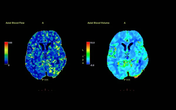
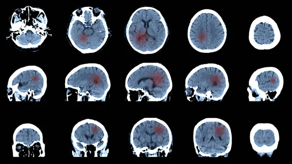
Stock image Axial view of CT Perusion of the brain with contrast showing brain anatomy, lobes, perfusion and function. The red area indicates high brain activity, perfusion and function.
Published:
Sep.26, 2022 11:28:56
Author:
samunella
Views:
12
Downloads:
1
File type:
image
/
jpg
File size:
2.32 MB
Orginal size:
2814
x
3096
px
Available sizes:
S
(454 x 500)
M
(909 x 1000)
L
(1818 x 2000)
XL
(2814 x 3096)
EL0
(2814 x 3096)
Level:
beginner
Download Image
Image topics:
Health
Head
Blood
Medicine
Medical
Technology
Flow
Doctor
Patient
Disease
Skull
Mind
Headache
Diagnosis
Stroke
Anatomy
Imaging
Surgery
Scan
Resonance
Advanced
Radiology
Artery
Cerebral
Neurology
Tumor
Lobe
Vascular
Infarction
Cerebrum
Hemorrhage
Tomography
Aneurysm
Perfusion
Computed
Medical Equipment
3d Rendering
Computed Tomography
Ct Scan
Blood Flow
Brain Scan
Apoplexy
CT Scanner
Cerebrovascular
Brain Tumors
Contrast Media
3d Reconstruction
Brain Perfusion
Perfusion Ct
Ct Brain Perfusion
Similar stock images
Colorized MRI Image Of Head Showing Brain
5200 × 3456
Perfusion CT Scan Of The Brain ,The Red And Green And Blue Are Areas Of Delayed Blood Flow To The Brain On The Perfusion CT Scan On The Screen.
3933 × 2476
Dorsal Striatum And Lateral Ventricles In The Brain Of A Person With Huntington's Disease, 3D Illustration Showing Enlargement Of Anterior Horns Of Lateral Ventricles And Atrophy Of The Caudate Nuclei
6000 × 4000
Brain Dorsal Striatum Anatomy, 3D Illustration. The Dorsal Striatum Consists Of The Caudate Nucleus (orange) And The Putamen (yellow). Lateral Ventricle Is Colored Blue, Amygdala Is Green
6000 × 4000
MRI Scan Brain, Ct Scan Of Brain Sagittal, Coronal, Transverse Image For Diagnosis
5999 × 3389