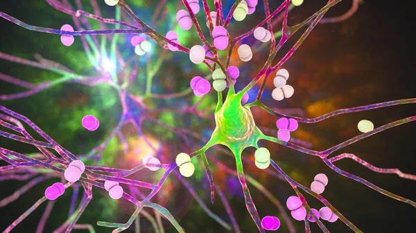
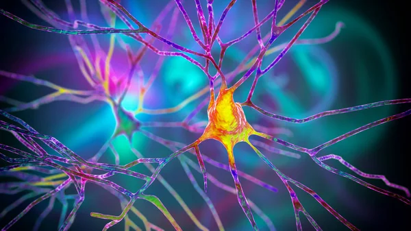
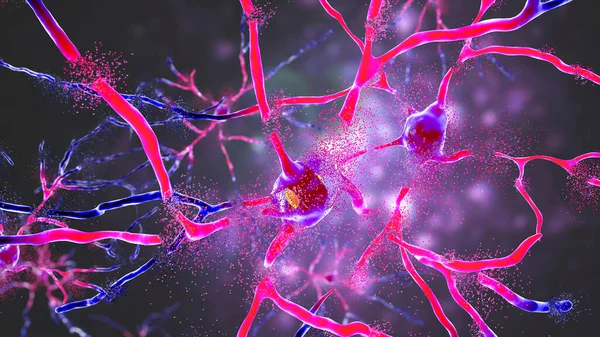
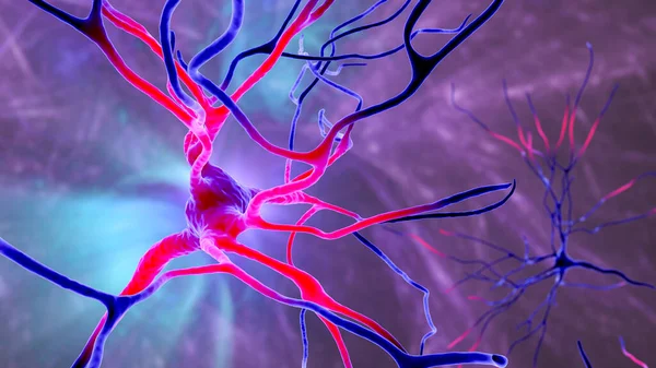
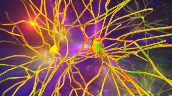
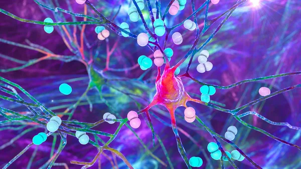
Stock image Bacteria Streptococcus pneumoniae infecting neurons, brain cells. Conceptual 3D illustration of pneumococcal and other bacterial encephalitis, meningitis, bacterial infection of brain tissue
Published: Aug.04, 2020 07:49:35
Author: katerynakon
Views: 5
Downloads: 0
File type: image / jpg
File size: 9.36 MB
Orginal size: 7200 x 4050 px
Available sizes:
Level: silver