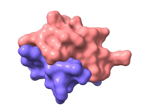
Stock image Bovine insulin, 3D-model of the heterodimer quaternary structure, chains A and B, surface filled model, white background
Published: Jun.23, 2020 06:42:42
Author: unnaugan
Views: 4
Downloads: 0
File type: image / jpg
File size: 0.71 MB
Orginal size: 4507 x 3380 px
Available sizes:
Level: beginner