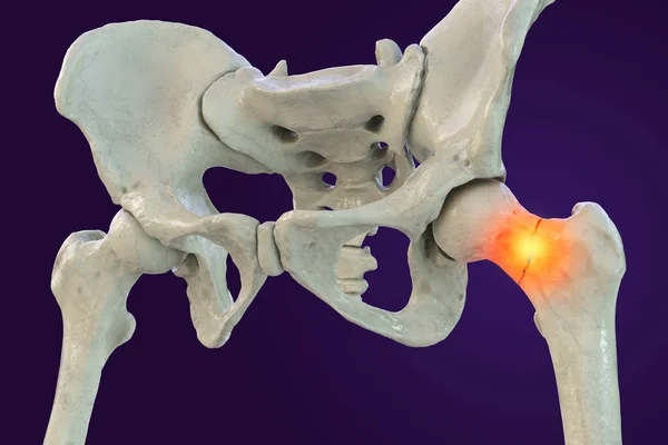
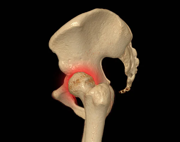
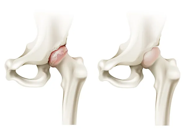
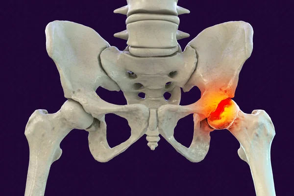
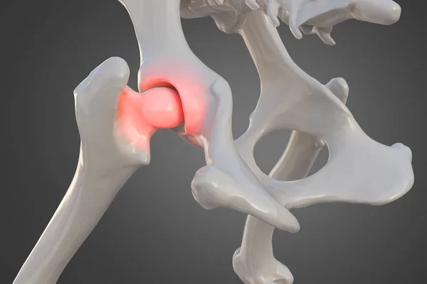
Stock image Canine dysplasia, dog bone with visible hip joint and femur affected by inflammation due to dysplasia, 3d illustration
Published: Jan.21, 2021 14:41:14
Author: Amaviael
Views: 21
Downloads: 1
File type: image / jpg
File size: 7.31 MB
Orginal size: 6000 x 4000 px
Available sizes:
Level: silver