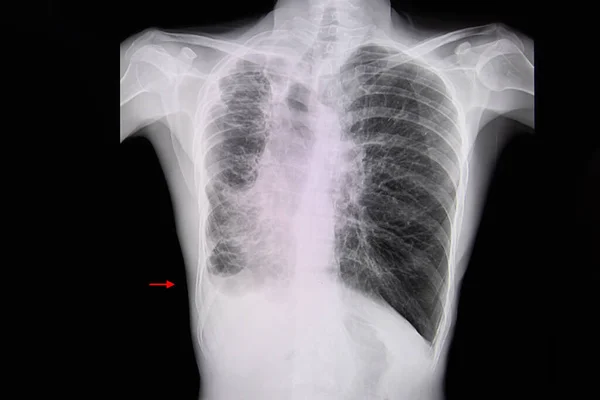
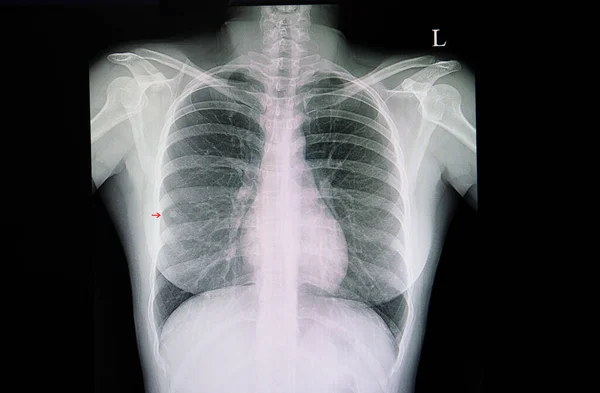
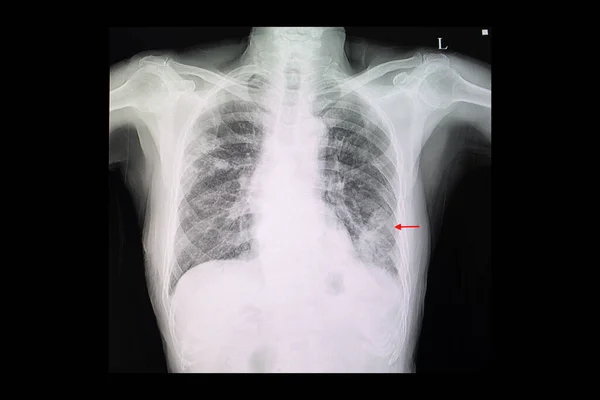
Stock image Chest x-ray of a patient with pneumonia of left lower lung with minimal pleural effusion.
Published: Sep.03, 2021 15:20:44
Author: navuths@gmail.com
Views: 42
Downloads: 0
File type: image / jpg
File size: 32.95 MB
Orginal size: 8192 x 5464 px
Available sizes:
Level: beginner
Similar stock images
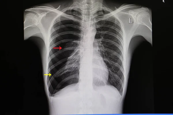
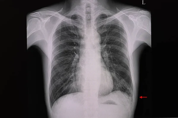
A Chest X-ray Film Of A Patient With Left Lower Lung Pneumonia And Right Upper Lung Nodules
6720 × 4480