Home
Images
Vectors
Videos
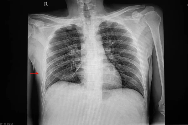
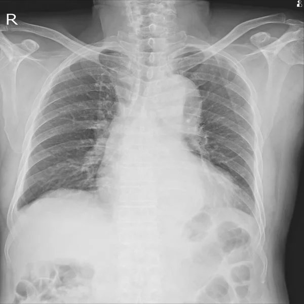
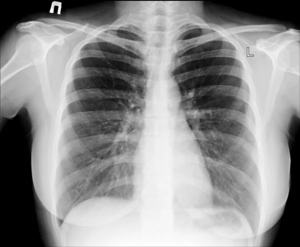
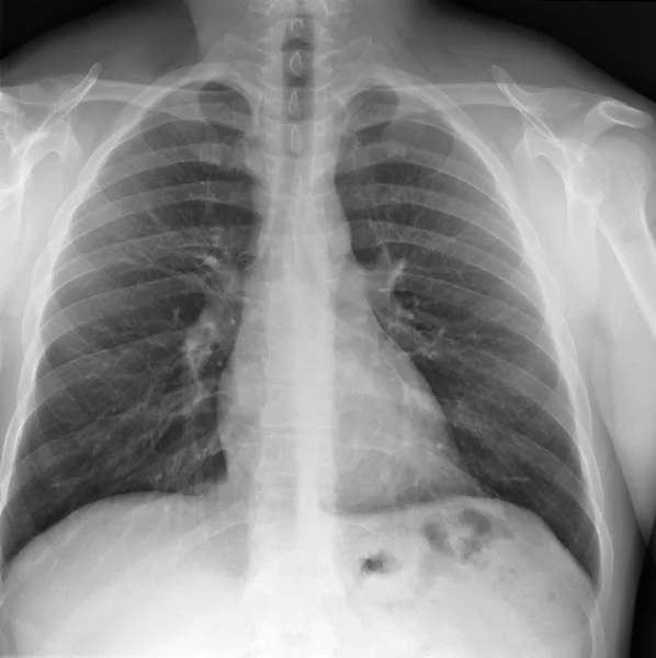
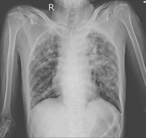
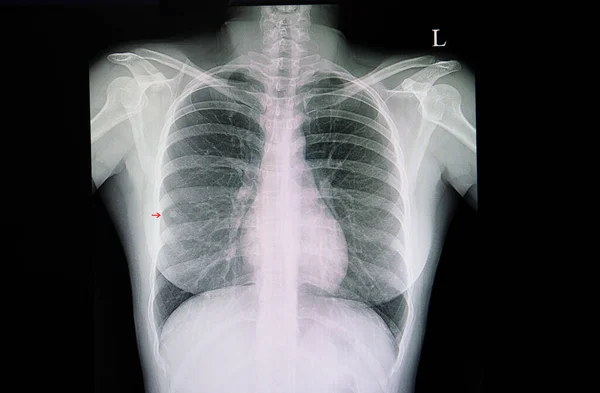
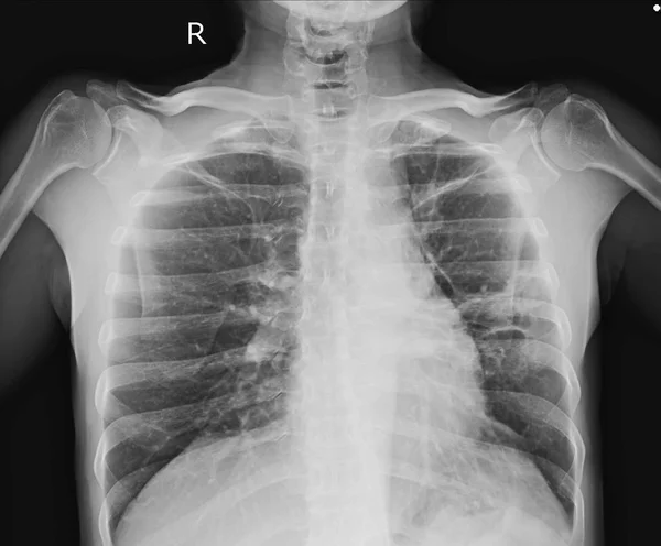
Stock image Chest x-ray showing A 3 cm pleural based nodule at Lt upper lobe with splicated margin. Thickening of nearby pleural. Nodular infiltration at RUL. Normal heart size. differential diagnosis CA lung, TB granuloma.
Published:
Feb.04, 2019 14:37:00
Author:
Richmanphoto
Views:
395
Downloads:
6
File type:
image
/
jpg
File size:
2.09 MB
Orginal size:
3628
x
3000
px
Available sizes:
S
(500 x 413)
M
(1000 x 827)
L
(2000 x 1654)
XL
(3628 x 3000)
EL0
(3628 x 3000)
Level:
bronze
Download Image
Image topics:
Image
Human
People
Health
Asthma
Medicine
Healthcare
Medical
Care
Doctor
Hospital
Bone
Stethoscope
Patient
Examining
Test
Body
Disease
Cancer
Lung
Film
Chest
Worker
Rib
Skeleton
Checkup
Diagnose
Diagnosis
Examination
Physician
Anatomy
Mucus
X Ray
Clinic
Scan
Surgeon
Medic
Specialist
Radiologist
Radiology
Xray
Bronchitis
Radiography
Pneumonia
Thorax
Pulmonary
Oncologist
Embolism
Cancerologist
Similar stock images
Chest X-ray Showing Small Pneumothorax On The Right Side Of Lung. The Pathology Causes By A Small Bronchopleural Fistula.
6157 × 4104
X-ray Chest Showing.Severe Cardiomegaly. Both Costophrenic Angles Are Clear.Intact Osseous StructureSevere Cardiomegaly.Infiltration At Lt Lower DDx. Pneumonia, Pulmonary Edema
3800 × 3800
X-ray Of Chest
2432 × 2004
X-ray Image Of Lung With Pneumonia.
2032 × 1964
X-ray Of Chest
2788 × 2796
Chest X-ray Showing Interstitial Pulmonary Infiltration With Dry Cavities Both Lungs.Impression : Pulmonary Tuberculosis
4028 × 3800
X-ray Image Of Lung With Pneumonia.
2032 × 1964
A Chest Xray Film Of A Patient With A Small Right Middle Lung Nodule.
6036 × 3959