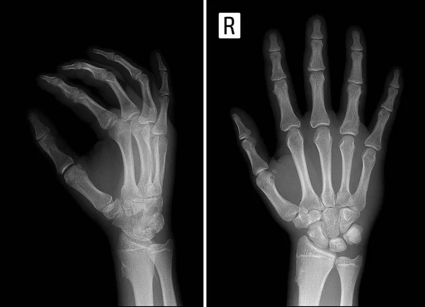
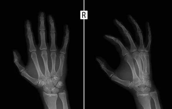
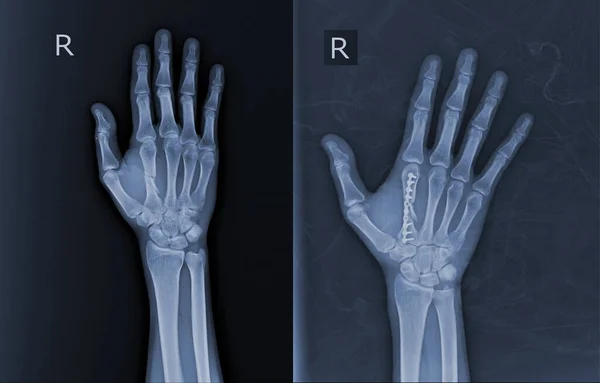
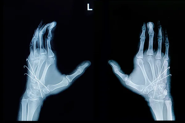
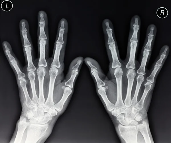
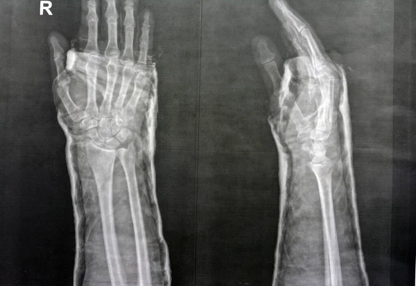
Stock image collection of x-ray image, hand x-ray AP and laterl view

Published: Dec.28, 2018 08:37:01
Author: Tridsanu
Views: 6
Downloads: 0
File type: image / jpg
File size: 2.12 MB
Orginal size: 2718 x 2947 px
Available sizes:
Level: bronze