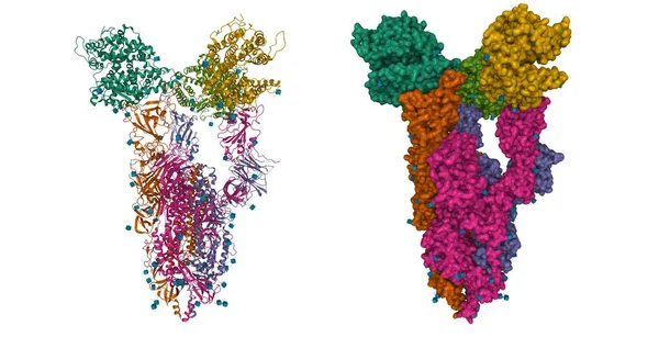
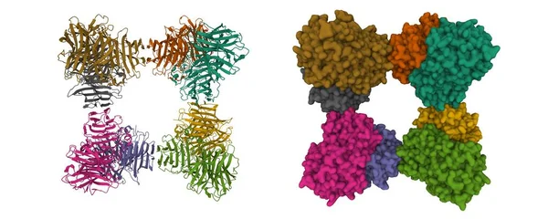
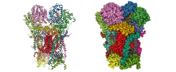
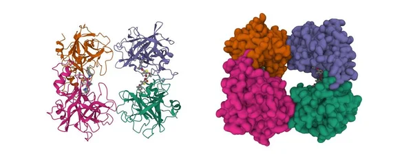
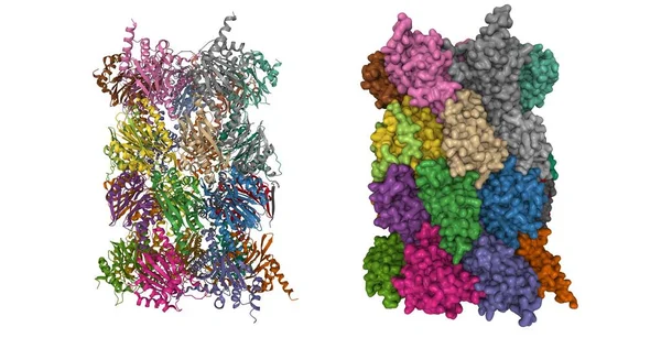
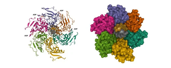
Stock image Cryo-EM structure of PCV2 replicase bound to ssDNA. 3D cartoon and Gaussian surface models, PDB 7las, chain id color scheme, white background
Published: Nov.08, 2022 14:50:09
Author: unnaugan
Views: 1
Downloads: 0
File type: image / jpg
File size: 5.69 MB
Orginal size: 10000 x 4000 px
Available sizes:
Level: beginner