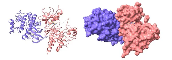
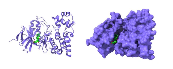
Stock image Crystal structure of human P450 3A4 in complex with erythromycin (red). The protoporphyrin is shown in green. 3D cartoon and Gaussian surface models, PDB 2j0D, white background.

Published: Jun.02, 2022 14:40:58
Author: unnaugan
Views: 4
Downloads: 0
File type: image / jpg
File size: 4.2 MB
Orginal size: 8000 x 4000 px
Available sizes:
Level: beginner