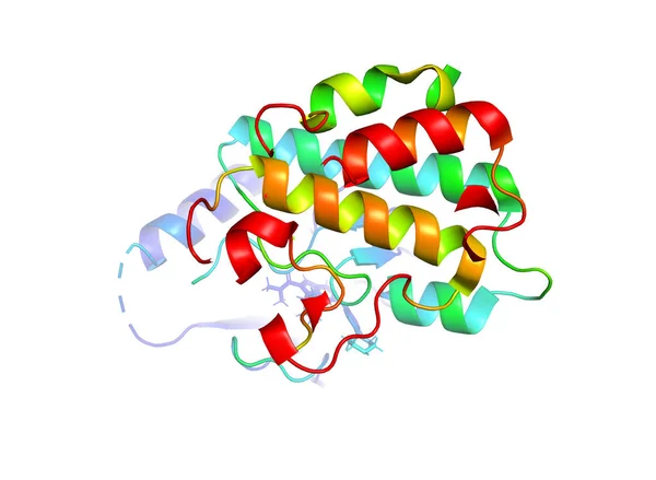
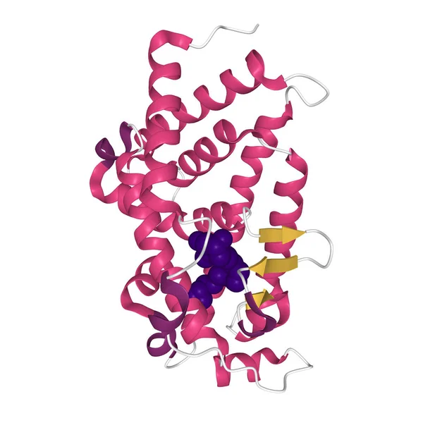
Stock image Crystal structure of VDR ligand binding domain complexed to calcipotriol (blue), 3D ball-and-stick model, white background
Published: Oct.05, 2020 07:00:06
Author: unnaugan
Views: 9
Downloads: 1
File type: image / jpg
File size: 2.54 MB
Orginal size: 4096 x 4096 px
Available sizes:
Level: beginner