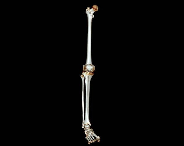
Stock image CT scan of lower extremity 3D for diagnosis fracture of Femur bone , knee joint , leg and foot 3D rendering

Published: Sep.12, 2022 08:14:15
Author: samunella
Views: 45
Downloads: 0
File type: image / jpg
File size: 1.55 MB
Orginal size: 3840 x 3036 px
Available sizes:
Level: beginner
Similar stock images
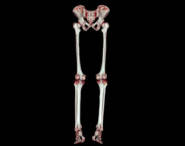
CT Scan Of Lower Extremity 3D For Diagnosis Fracture Of Femur Bone , Knee Joint , Leg And Foot 3D Rendering
3840 × 3036
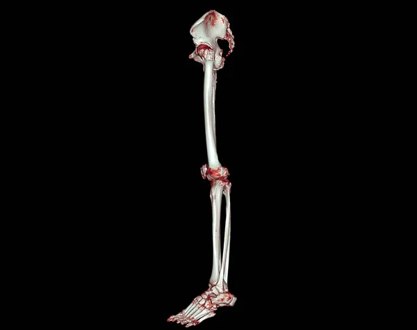
CT Scan Of Lower Extremity 3D For Diagnosis Fracture Of Femur Bone , Knee Joint , Leg And Foot 3D Rendering
3840 × 3036