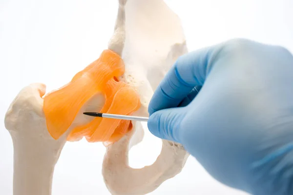
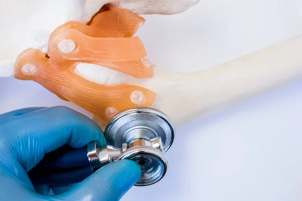
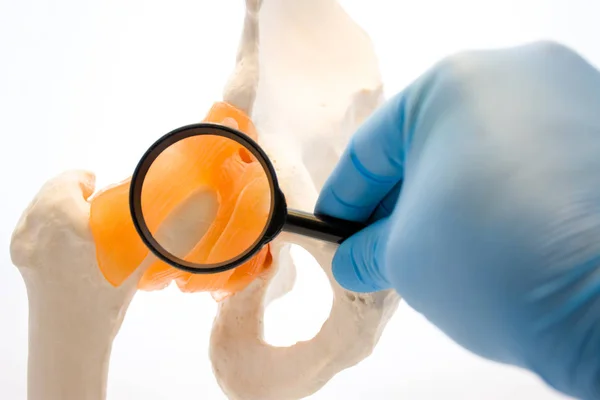
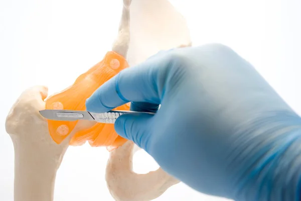
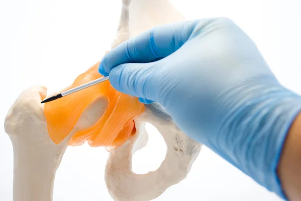
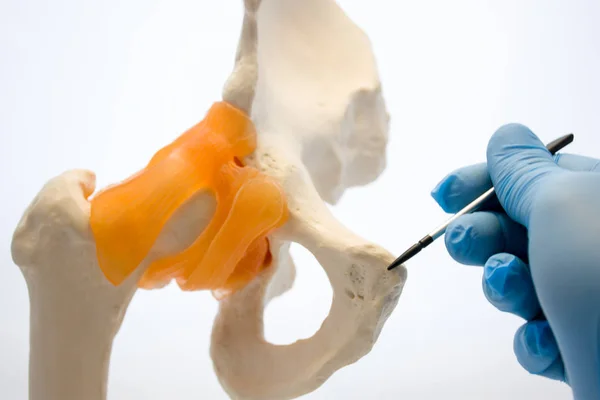
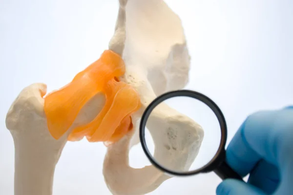
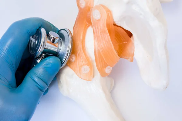
Stock image Diagnosis of diseases of joints and bones concept photo. Doctor checks or examine by stethoscope hip joint and femur on anatomical model. Visualization of joints and bones diagnostics in orthopedics
Published: Dec.14, 2017 16:22:13
Author: Shidlovski
Views: 47
Downloads: 1
File type: image / jpg
File size: 6.9 MB
Orginal size: 6000 x 4000 px
Available sizes:
Level: bronze