Home
Images
Vectors
Videos
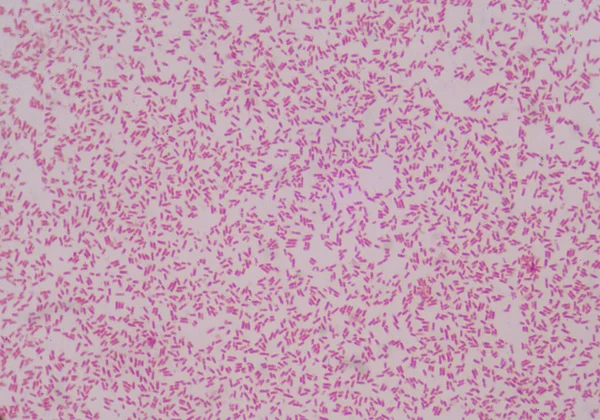
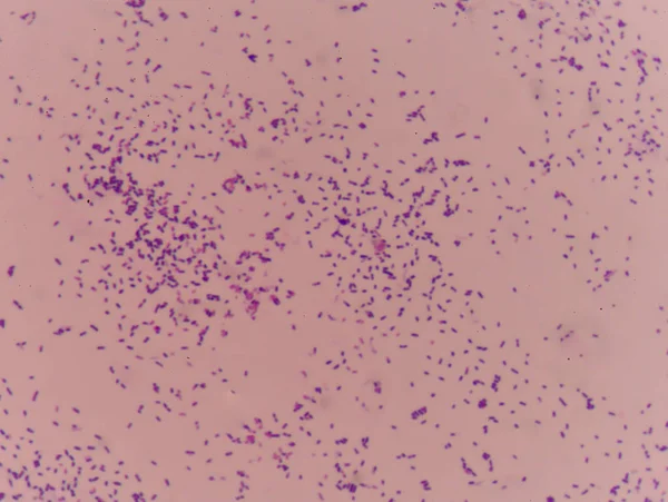
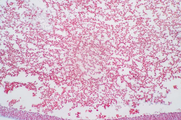
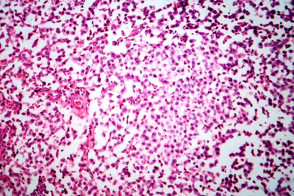
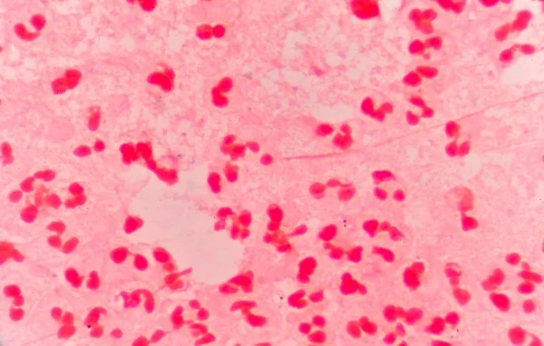
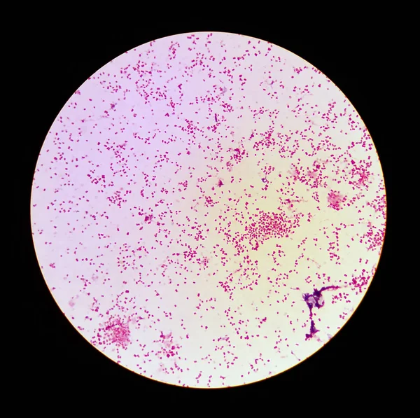
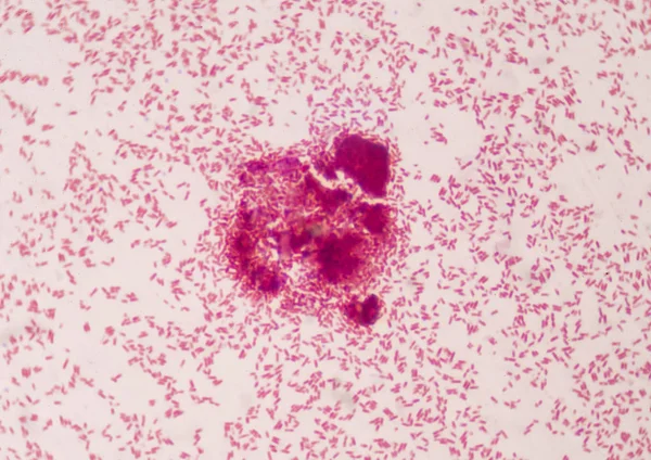
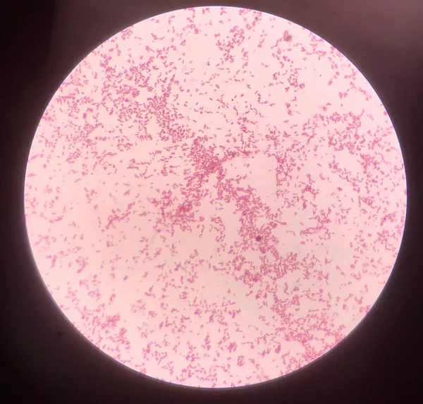
Stock image Gram staining, also called Gram's method, is a method of differentiating bacterial species into two large groups (Gram-positive and Gram-negative).
Published:
Nov.01, 2018 12:17:48
Author:
toeytoey
Views:
713
Downloads:
17
File type:
image
/
jpg
File size:
15.15 MB
Orginal size:
4998
x
3594
px
Available sizes:
S
(500 x 360)
M
(1000 x 719)
L
(2000 x 1438)
XL
(4998 x 3594)
EL0
(4998 x 3594)
Level:
bronze
Download Image
Image topics:
Red
Shape
Human
Health
Medicine
Healthcare
Medical
Care
Wall
Stain
Acid
Bacterial
Cell
Organic
Disease
Biology
Lab
Laboratory
Science
Fast
Negative
Stains
Bacterium
Positive
Microorganism
Microbe
Rod
Gram
Bacteria
Microscope
Dna
Microbiology
Method
Outbreak
Germs
Infection
Staining
Bacilli
Plasmid
Flagella
Cocci
Similar stock images
Moderate Grem Neagtive Bacilli Medicl Science Co Cept.
4893 × 3428
Gram Stain Test Showing Bacteria Cells.
4695 × 3536
Anatomy And Histological Ovary And Testis Human Cells Under Microscope.
6720 × 4480
Testicular Seminoma, Light Micrograph, Photo Under Microscope. A Most Common Germ Cell Tumor Of The Testis
4434 × 2956
Moderate Red White Blood Cells With Gram Negative Diplococci.
5455 × 3475
Microscopic View Of Gonorrhea (Neisseria Gonorrhoeae)
2309 × 2300
Bacteria Intracellular Red Cells In Gramstain Find With Microscope.
4987 × 3526
Bacteria Cell In Gram Stain.
3162 × 3024