Home
Images
Vectors
Videos
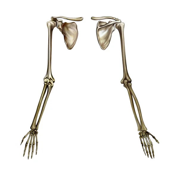
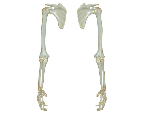
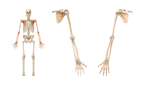
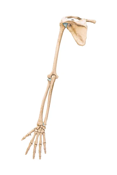
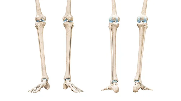
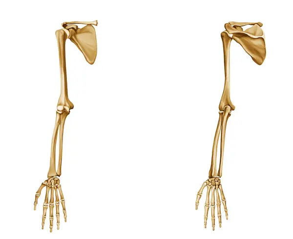
Stock image Hand Bone Anatomy Front & Back White background
Published:
May.18, 2024 18:10:21
Author:
vishmaya88@gmail.com
Views:
1
Downloads:
0
File type:
image
/
jpg
File size:
3.27 MB
Orginal size:
6120
x
5000
px
Available sizes:
S
(500 x 408)
M
(1000 x 817)
L
(2000 x 1634)
XL
(6120 x 5000)
EL0
(6120 x 5000)
Level:
beginner
Download Image
Image topics:
Lunate
Trapezium
Trapezoid
Capitate
Triquetrum
Pisiform
Hamate
Phalanges
Dorsal View
Carpals
Metacarpals
Proximal Phalanges
Middle Phalanges
Palmar View
Similar stock images
Human Hand Anatomy, Bones Of The Shoulder Girdle, Shoulder And Forearm, Realistic Drawing, Skeleton Structure, Front And Back View, Isolated Image On A White Background
3356 × 3425
Human Skeleton Isolated On White Background
4800 × 3200
CT Scan Of Lower Extremity 3D For Diagnosis Fracture Of Femur Bone , Knee Joint , Leg And Foot 3D Rendering
3840 × 3036
Leg Bones (Membrum Inferius Liberum), Realistic Drawing Of The Human Skeleton, Front And Back View, Femur, Tibia, Knee, Patella, Metatarsus, Foot Structure, Anatomy, Body Parts, Isolated Image On A White Background
2608 × 3425
CT SCAN Of Upper Extremity 3D Rendering Isolated On White Background .Clipping Path.
3840 × 3036
Accurate Bones Of The Arm Or Upper Limb Of The Human Skeletal System Or Skeleton Isolated On White Background 3D Rendering Illustration. Anterior And Posterior Views. Anatomy, Osteology Concept.
3800 × 2271
Accurate Posterior Or Rear View Of The Arm Or Upper Limb Bones Of The Human Skeletal System Isolated On White Background 3D Rendering Illustration. Anatomy, Medical, Osteology Concept.
2000 × 3000
Tibia Or Shin Bone Front And Rear Views 3D Rendering Illustration Isolated On White With Copy Space. Human Skeleton And Leg Anatomy, Medical Diagram, Osteology, Skeletal System Concepts.
4484 × 2500