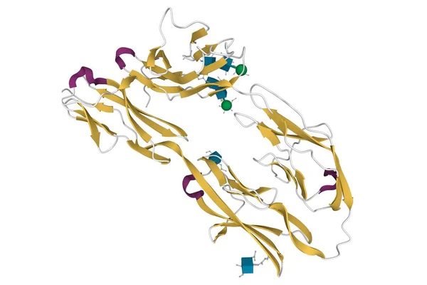
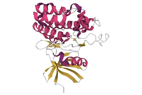
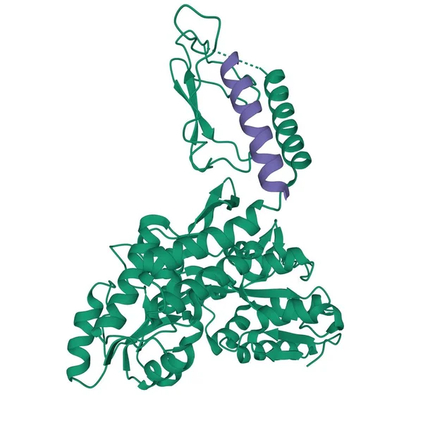
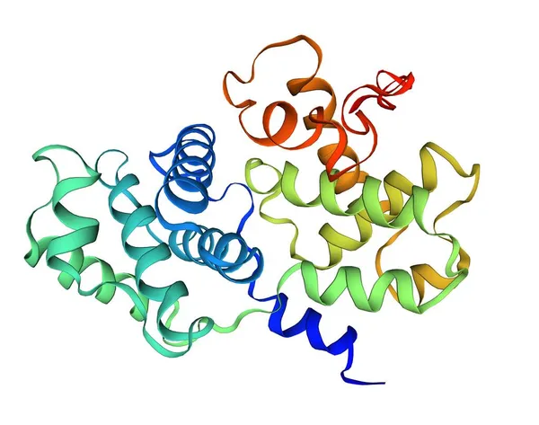
Stock image Human ACE2 receptor, 3D illustration. Angiotensin Converting Enzyme-Related Carboxypeptidase, a membrane protein which is used by SARS-CoV-2 virus to enter the human cells

Published: Jul.19, 2021 08:47:03
Author: katerynakon
Views: 0
Downloads: 2
File type: image / jpg
File size: 4.71 MB
Orginal size: 6000 x 4000 px
Available sizes:
Level: silver