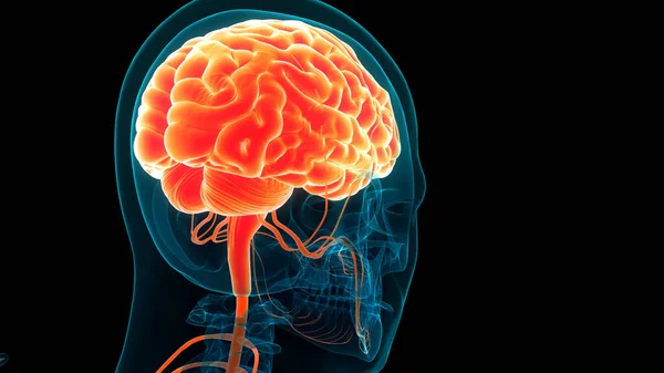
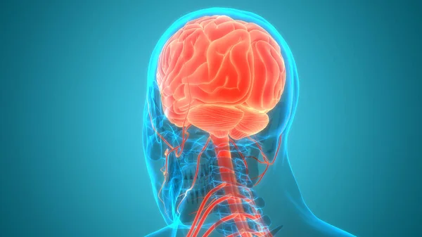
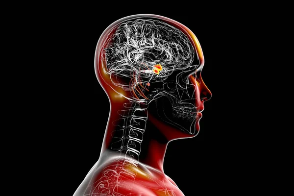
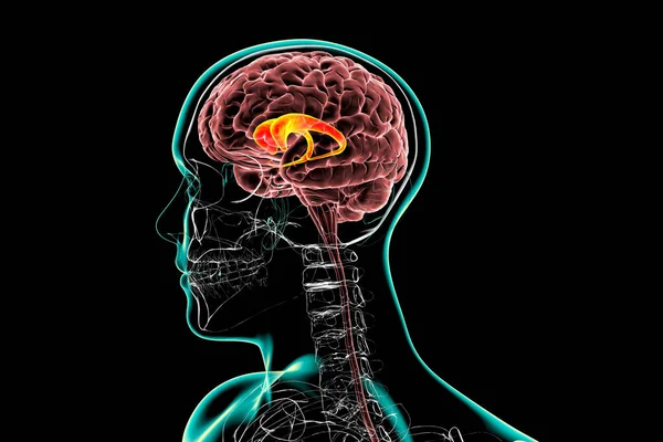
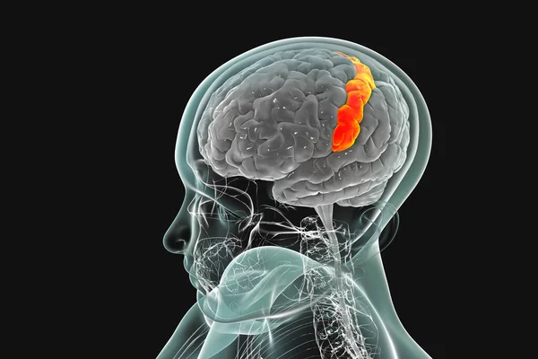
Stock image Human brain in body with highlighted postcentral gyrus, 3D illustration. It is located in the lateral parietal lobe, the primary somatosensory cortex, and is responsible for the sense of touch
Published: Jan.14, 2021 09:08:43
Author: katerynakon
Views: 57
Downloads: 2
File type: image / jpg
File size: 4.19 MB
Orginal size: 6000 x 4000 px
Available sizes:
Level: silver