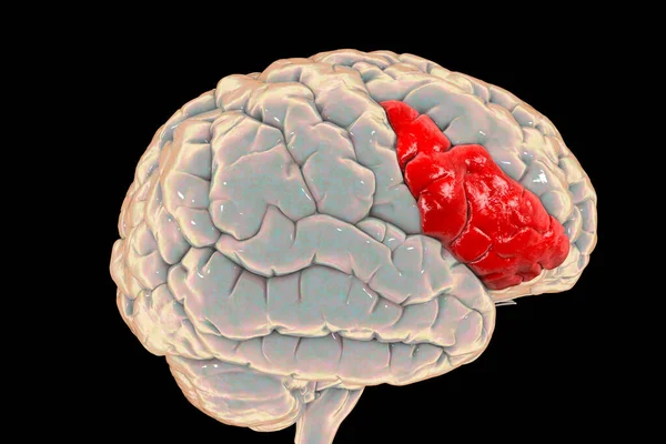
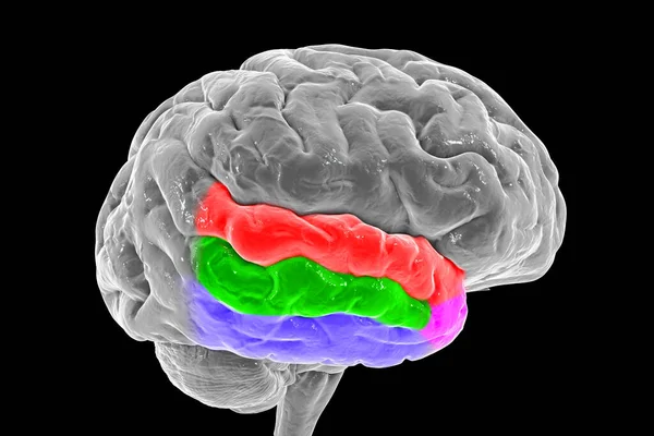
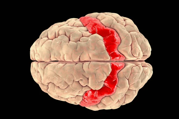
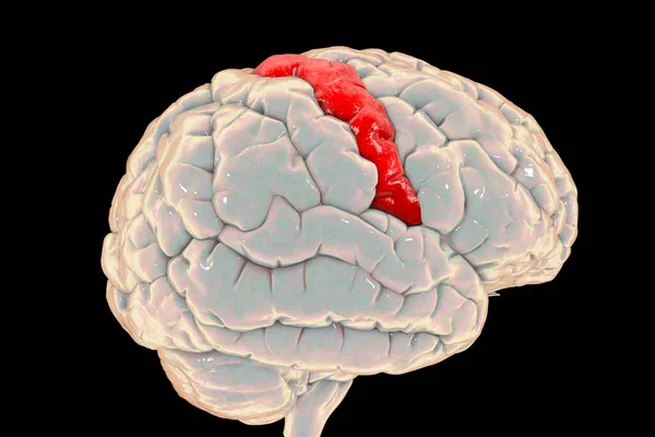
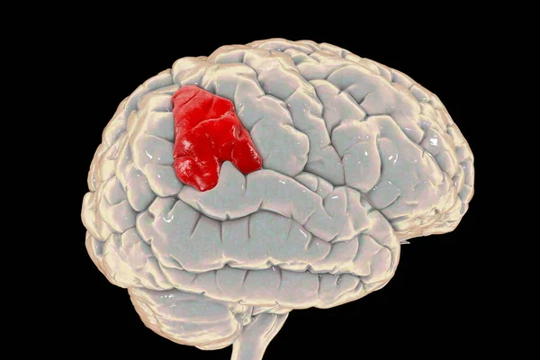
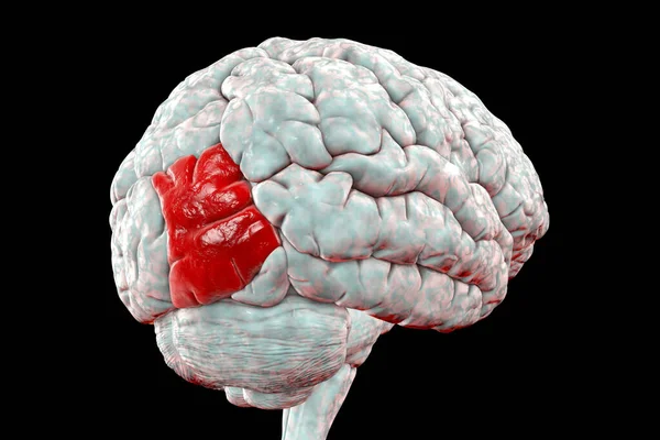
Stock image Human brain with highlighted middle (lateral) occipital gyrus, 3D illustration. It is located in occipital lobe and is responsible for object recognition
Published: Dec.01, 2020 16:21:16
Author: katerynakon
Views: 20
Downloads: 0
File type: image / jpg
File size: 5.52 MB
Orginal size: 6202 x 4135 px
Available sizes:
Level: silver