Home
Images
Vectors
Videos
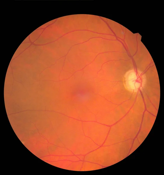
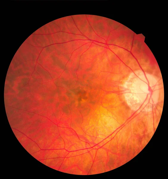
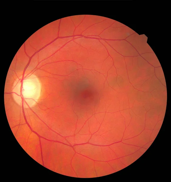
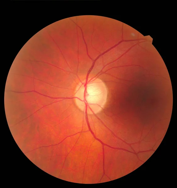
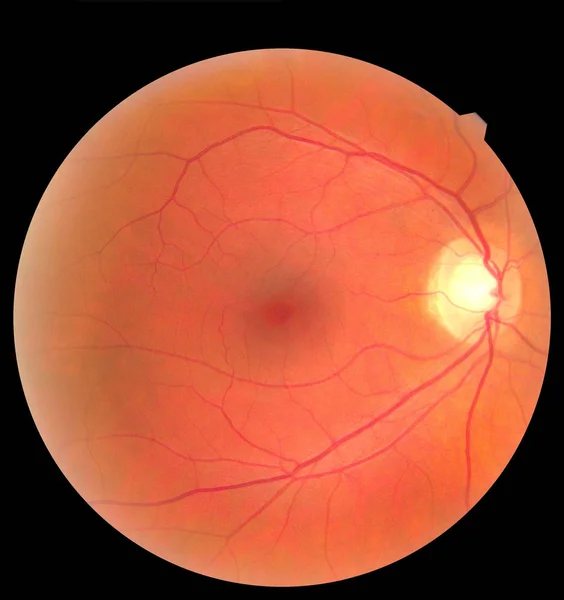
Stock image Human eye anatomy taking images with Mydriatic Retinal cameras. Examination of the eye, diabetic retinopathy, ARMD
Published:
Apr.23, 2020 10:40:43
Author:
ternavskaia.o@gmail.com
Views:
30
Downloads:
6
File type:
image
/
jpg
File size:
2.98 MB
Orginal size:
3000
x
3186
px
Available sizes:
S
(471 x 500)
M
(942 x 1000)
L
(1883 x 2000)
XL
(3000 x 3186)
EL0
(3000 x 3186)
Level:
bronze
Download Image
Image topics:
Background
View
Macro
Focus
Isolated
Beautiful
Field
Light
Blood
Illness
Medical
Care
Eye
Lens
Hole
Vision
Ill
Vein
Optic
Iris
Disc
Anatomy
Ophthalmic
Ophthalmologist
Mild
Severe
Ocular
Specialist
Eyeball
Artery
Chamber
Diabetic
Vessels
Anatomical
Retina
Arterial
Retinal
Posterior
Hemorrhage
Oculus
Haemorrhage
Ischemic
Visualize
Dics
Fundus
Ischemia
Macula
Retinopathy
Ciliary
Armd
Similar stock images
Human Eye Anatomy Taking Images With Mydriatic Retinal Cameras. Examination Of The Eye, Diabetic Retinopathy, ARMD
3000 × 3186
Human Eye Anatomy Taking Images With Mydriatic Retinal Cameras. Examination Of The Eye, Diabetic Retinopathy, ARMD
3000 × 3186
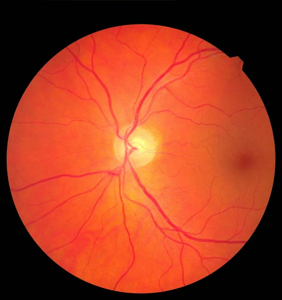
Ophthalmic Image Detailing The Retina And Optic Nerve Inside A Healthy Human Eye. Health Protection Concept
3000 × 3186
View Inside Human Eye Showing Retina, Optic Nerve And Macula. Health Concept
3000 × 3186
Ophthalmic Image Detailing The Retina And Optic Nerve Inside A Healthy Human Eye. Medicine Concept
3000 × 3186
View Inside Human Eye Showing Retina, Optic Nerve And Macula. Health Concept
3000 × 3186
Ophthalmic Image Detailing The Retina And Optic Nerve Inside A Healthy Human Eye. Medicine Concept
3000 × 3186
View Inside Human Eye Showing Retina, Optic Nerve And Macula. Health Concept
3000 × 3186