Home
Images
Vectors
Videos
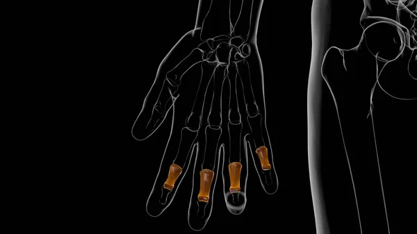
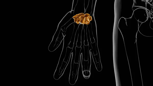
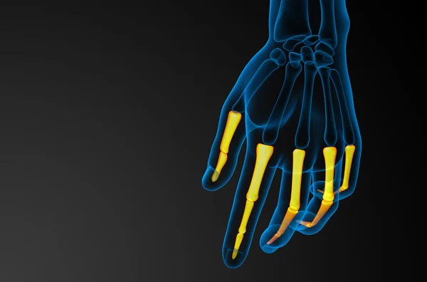
Stock image Human Skeleton Proximal Phalanges Anatomy 3D Illustration
Published:
Jun.02, 2021 10:16:15
Author:
My_box_pra
Views:
1
Downloads:
0
File type:
image
/
jpg
File size:
0.76 MB
Orginal size:
3840
x
2160
px
Available sizes:
S
(500 x 281)
M
(1000 x 563)
L
(2000 x 1125)
XL
(3840 x 2160)
EL0
(3840 x 2160)
Level:
bronze
Download Image
Image topics:
Skeleton
Anatomy
Temporal
Lunate
Thoracic
Maxilla
Cervical
Inferior
Trapezium
Trapezoid
Ethmoid
Capitate
Scaphoid
Pisiform
Hamate
Vomer
Septal
3d Rendering
Rib Cage
3d Illustration
Carpals
Manubrium
Metacarpals
Frontal Bone
Appendicular
Occipital Bone
Nasal Bone
Axial Skeleton
Lacrimal Bone
Sphenoid Bone
Palatine Bone
Triquetral
Sacrum Bone
Proximal Phalanges
Intermediate Phalanges
Xiphoid Process
Nasal Concha
Distal Phalanges
Clavicle Bone
Nasal Cartilage
Similar stock images
Human Skeleton Hand Distal Phalanges Bone Anatomy For Medical Concept 3D Illustration
3840 × 2160
Human Skeleton Intermediate Phalanges Anatomy 3D Illustration
3840 × 2160
Human Skeleton Hand Matacarapls Bone Anatomy For Medical Concept 3D Illustration
3840 × 2160
Human Skeleton Hand Wrist Carpals Bone Anatomy For Medical Concept 3D Illustration
3840 × 2160
X-RAY Skeleton 3d Illustration With Copy Space
5940 × 4200
Transparent Female Skeleton - Hand Bones
3750 × 5000
3d Render Illustration Of The Human Phalanges Hand
4649 × 3072
Muscle Is A Soft Tissue, Muscle Cells Contain Proteins , Producing A Contraction That Changes Both The Length And The Shape Of The Cell. Muscles Function To Produce Force And Motion.
4096 × 4096