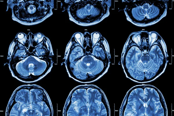
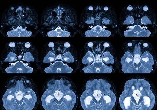
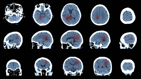
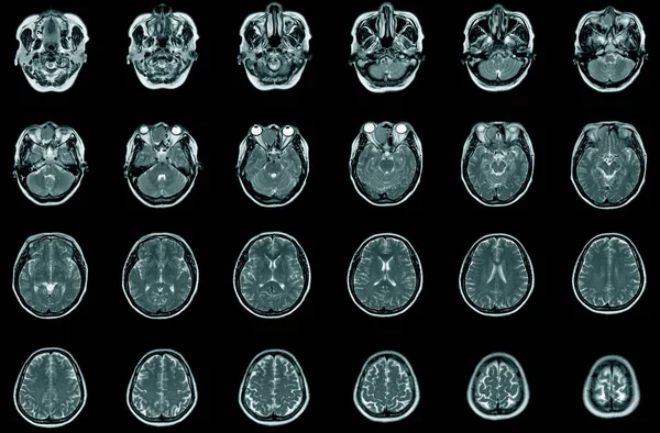
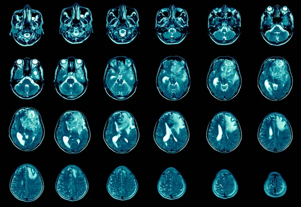
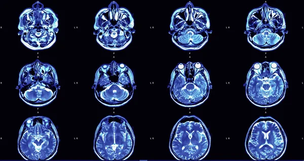
Stock image magnetic resonance image (MRI) of the brain
Published: Sep.23, 2019 07:17:03
Author: akesin@gmail.com
Views: 12
Downloads: 0
File type: image / jpg
File size: 6.94 MB
Orginal size: 6000 x 3194 px
Available sizes:
Level: bronze