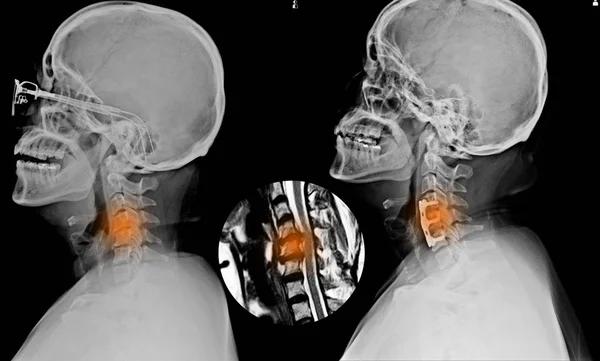
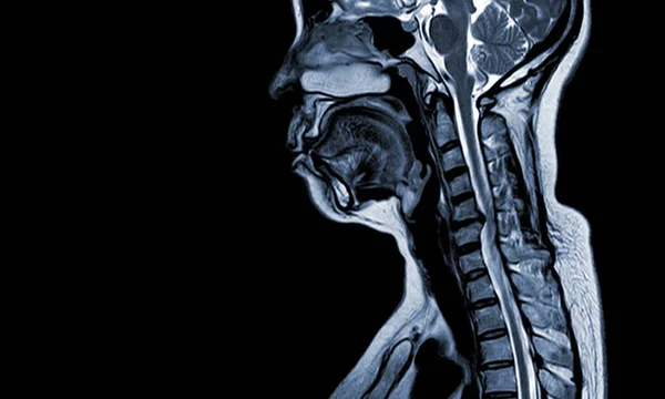
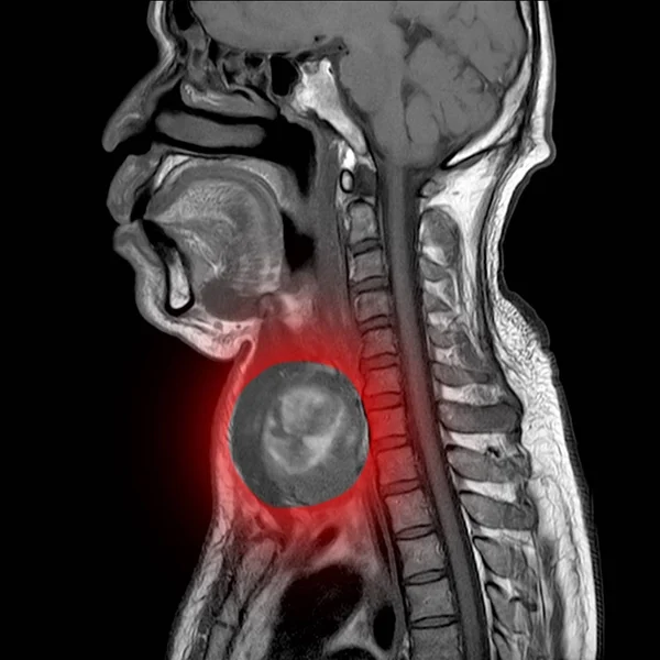
Stock image Magnetic resonance imaging (MRI) of neck,
Published: Dec.06, 2019 07:17:25
Author: Richmanphoto
Views: 326
Downloads: 1
File type: image / jpg
File size: 2.23 MB
Orginal size: 3500 x 3500 px
Available sizes:
Level: bronze