Home
Images
Vectors
Videos
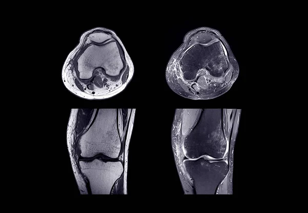
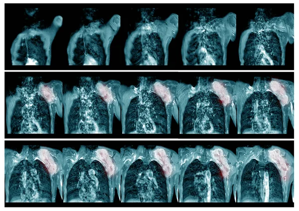
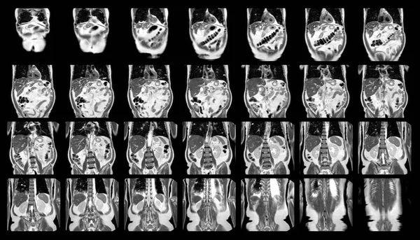
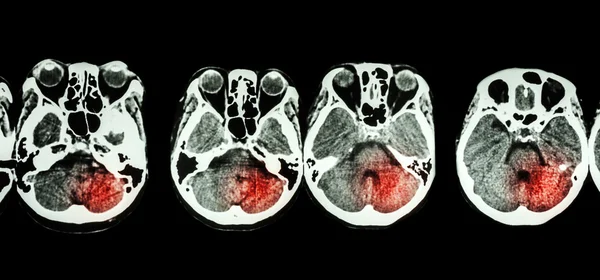
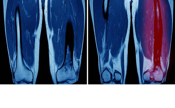
Stock image magnetic resonance imaging( MRI )of right thigh Findings:fat containing mass in vastus intermedius muscle.shows heterogeneous enhancement predominate at superomedial aspect of the mass, suggestive of recurrent liposarcoma
Published:
Mar.30, 2020 06:10:46
Author:
Richmanphoto
Views:
97
Downloads:
0
File type:
image
/
jpg
File size:
5.85 MB
Orginal size:
5788
x
3500
px
Available sizes:
S
(500 x 302)
M
(1000 x 605)
L
(2000 x 1209)
XL
(5788 x 3500)
EL0
(5788 x 3500)
Level:
bronze
Download Image
Image topics:
Sport
Human
People
Health
Medicine
Medical
Technology
Doctor
Hospital
Bone
Patient
Body
Therapy
Broken
Film
Ray
Leg
Vessel
Pain
Knee
Sick
Muscle
Joint
Structures
Magnetic
Skeleton
Magnet
Diagnosis
Examination
Anatomy
Limb
X Ray
Surgery
Clinic
Trauma
Injury
Scan
Diagnostic
Resonance
Scanner
Artery
Tumor
Orthopedic
Mri
Femoral
Femur
Cartilage
Effusion
Arthrosis
Condyle
Similar stock images
MRI Of Right Knee
3149 × 2173
Magnetic Resonance Imaging(MRI) Left.shoulder History:Case Lt. Shoulder Mass . Impression:-The Mass Soft Tissue Sarcoma And Metastasis.Medical And Healthcare Concept.
8552 × 6068
Magnetic Resonance Imaging (MRI)OF UPPER ABDOMEN Of A Patient Who Has Suspected Mass-forming Hilar Cholangiocarcinoma Coronal View Science And Education.Medical Technology And Science Concept
6240 × 3580
Stroke ( CT Scan Of Brain And Base Of Skull And Stroke )
5127 × 2392
MRI Spine Showing Mass At Distal Cord And Conus Medullaris, With Tumor Seedings At Lower Spinal Canal. The Mass Appear Hypersignal T2 And Strong Enhancement. DDx. Myxopapillary Ependymoma.
6564 × 3940
Magnetic Resonance Imaging Pelvic Of A Healthy Woman Finding Abnormalities Like Mass Lumps.Medical Healthcare Concept.
6000 × 4000
Set Of MRI Scanner Slices Of Dental Part Of Human Male Scull With Multiple Problems On White Background.
3588 × 2041
Magnetic Resonance Imaging (MRI) Of Both Femur.Impression : Chronic Osteomyelitis Left Femur On Red Mark. And Copo Space ,Medical Healthcare Concept
4672 × 2888