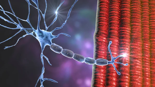
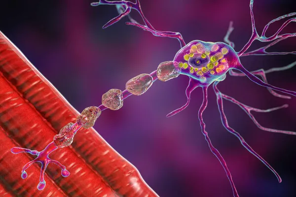
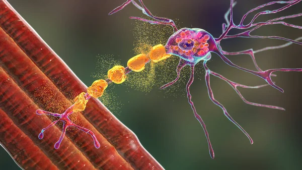
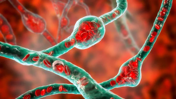
Stock image Motor neuron connecting to muscle fiber, 3D illustration. A neuromuscular junction allows the motor neuron to transmit a signal to the muscle causing contraction. It is affected by toxins and diseases

Published: Apr.27, 2021 09:28:02
Author: katerynakon
Views: 37
Downloads: 3
File type: image / jpg
File size: 15.53 MB
Orginal size: 6075 x 4050 px
Available sizes:
Level: silver