Home
Images
Vectors
Videos
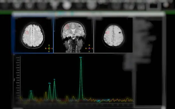
Stock image MR spectroscopy aids in stroke diseases, providing insightful chemical analysis to understand metabolic changes in affected brain tissues.
Published:
May.02, 2024 08:48:14
Author:
samunella
Views:
0
Downloads:
0
File type:
image
/
jpg
File size:
1.22 MB
Orginal size:
3456
x
2160
px
Available sizes:
S
(500 x 313)
M
(1000 x 625)
L
(2000 x 1250)
XL
(3456 x 2160)
EL0
(3456 x 2160)
Level:
beginner
Download Image
Image topics:
Emergency
Intensive
Anaesthesia
Diagnostic Tool
Spectroscopy
Chemical Analysis
Life Support
Neurological Disorders
Healing Process
Non Invasive Method
Mri Brain
Stroke Patient
Cellular Metabolism
Treatment Response
Neurological Assessment
MRI Brain Scan
Brain Tissues
Cellular Health
Precision Medicine
Molecular Level
Clinical Decision Making
Neurological Research
Therapeutic Strategies
Advanced Imaging
Insightful Analysis
Stroke Diagnosis
Biochemical Markers
Mr Spectroscopy
Lesion Metabolism
Lesion Analysis
Metabolic Changes
Mri Brain Spectroscopy
Disease Monitoring
Spectroscopic Examination
Biochemical Changes
Stroke Diseases
Treatment Assessment
Metabolic Profiling
Neurological Imaging
Molecular Insights
Stroke Cases
Non Destructive Analysis
Metabolite Concentrations
Neurochemical Evaluation
Clinical Application
Similar stock images
MR Spectroscopy Aids In Stroke Diseases, Providing Insightful Chemical Analysis To Understand Metabolic Changes In Affected Brain Tissues.
3456 × 2160
Close-up Of An MRI Of The Brain And Blood Vessels Of A Person With Severe Headaches; Magnetic And Nuclear Resonance And Angiography As A Diagnostic Method In Neurology
4704 × 2169
MR Spectroscopy Aids In Stroke Diseases, Providing Insightful Chemical Analysis To Understand Metabolic Changes In Affected Brain Tissues.
4000 × 3000
MRI Or Magnetic Resonance Image Of Head And Brain Scan. Close Up View
3001 × 2001
CT Scan Of The Brain Axial, Coronal And Sagittal View For Diagnosis Brain Tumor,stroke Diseases And Vascular Diseases.
3840 × 2400
MRI Brain Scan With Problems
3200 × 2016
Images From A Computerized Tomography
4928 × 3280
CT And MRI Of The Skull
3900 × 2600