Stock image MRI of the upper abdomen is a non-invasive imaging technique providing detailed visuals of organs like the liver, pancreas, and kidneys in case normal study.

Published: Apr.27, 2024 04:06:18
Author: samunella
Views: 1
Downloads: 0
File type: image / jpg
File size: 3.15 MB
Orginal size: 5601 x 3258 px
Available sizes:
Level: beginner
Similar stock images
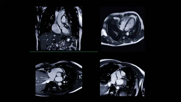
MRI Heart Or Cardiac MRI ( Magnetic Resonance Imaging ) Of Heart For Detecting Heart Disease.
5760 × 3240