Home
Images
Vectors
Videos
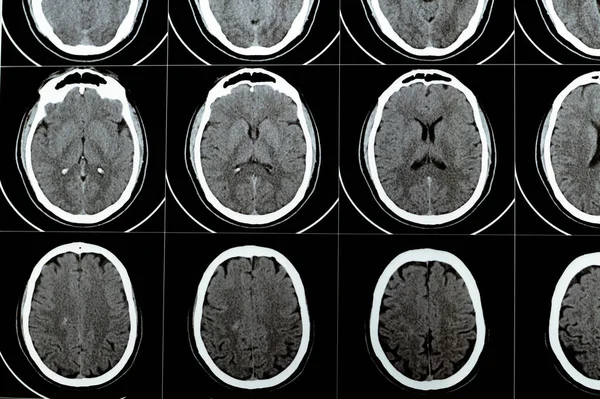
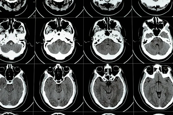
Stock image Multi slice CT scan of the brain showing Large brain stem and right centrum semiovale hematoma, normal posterior fossa structures, normal size of ventricular system and central mid-line structures
Published:
Aug.09, 2023 09:36:33
Author:
Tamer_Soliman
Views:
2
Downloads:
0
File type:
image
/
jpg
File size:
5.83 MB
Orginal size:
6016
x
4000
px
Available sizes:
S
(500 x 332)
M
(1000 x 665)
L
(2000 x 1330)
XL
(6016 x 4000)
EL0
(6016 x 4000)
Level:
beginner
Download Image
Image topics:
Image
Human
Health
Head
Medicine
Medical
Stem
Cat
Patient
Disease
Ray
Skull
Diagnosis
Examination
Hypertension
Stroke
Brain
X Ray
Accident
Scan
Diagnostic
Disorder
Radiologist
Radiology
Cerebral
Neurology
Vessels
Abnormal
Radiography
Vascular
Mri
Cerebrum
Medulla
Ct
Hemorrhage
Neurologist
Tomography
Hematoma
Haemorrhage
Hemorrhagic
Ischemic
Computed
Thrombosis
Oblongata
Cvs
Hemangioma
Cerebrovascular
Arteriovenous
Cavernoma
Similar stock images
Multi Slice CT Scan Of The Brain Showing Large Brain Stem And Right Centrum Semiovale Hematoma, Normal Posterior Fossa Structures, Normal Size Of Ventricular System And Central Mid-line Structures
6016 × 4000
Multi Slice CT Scan Of The Brain Showing Large Brain Stem And Right Centrum Semiovale Hematoma, Normal Posterior Fossa Structures, Normal Size Of Ventricular System And Central Mid-line Structures
6016 × 4000
Multi Slice CT Scan Of The Brain Showing Large Brain Stem And Right Centrum Semiovale Hematoma, Normal Posterior Fossa Structures, Normal Size Of Ventricular System And Central Mid-line Structures
5792 × 4000
Multi Slice CT Scan Of The Brain Showing Large Brain Stem And Right Centrum Semiovale Hematoma, Normal Posterior Fossa Structures, Normal Size Of Ventricular System And Central Mid-line Structures
6016 × 4000
Brain CT Scan Showing Brainstem Cavernoma, Right Centrum Semiovale Developmental Venous Anomaly, Intra Cerebral Haematoma, Faint Hypodense Lesion In Medulla Oblongata And Pontomedullary Junction
5890 × 3804
Multi Slice CT Scan Of The Brain Showing Large Brain Stem And Right Centrum Semiovale Hematoma, Normal Posterior Fossa Structures, Normal Size Of Ventricular System And Central Mid-line Structures
6016 × 4000
A CT Scan Image Of The Human Skull With Intracranial Shunt
4288 × 2848
Brain CT Scan Showing Brainstem Cavernoma, Right Centrum Semiovale Developmental Venous Anomaly, Intra Cerebral Haematoma, Faint Hypodense Lesion In Medulla Oblongata And Pontomedullary Junction
6016 × 4000