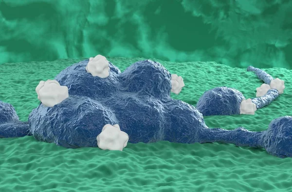
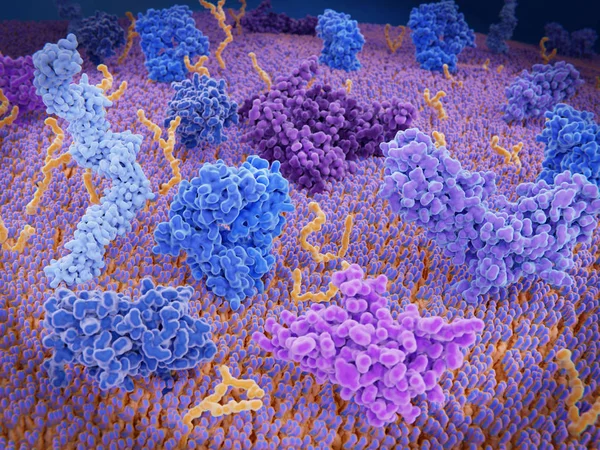
Stock image Neurotoxins (like spyder venom enzyme) destructive to nerve tissue - 3d illustration closeup view

Published: May.18, 2023 07:54:19
Author: forfour
Views: 2
Downloads: 0
File type: image / jpg
File size: 9.87 MB
Orginal size: 10000 x 6600 px
Available sizes:
Level: beginner