Home
Images
Vectors
Videos
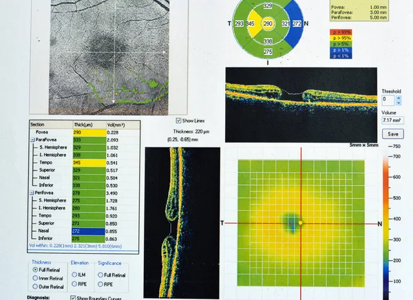
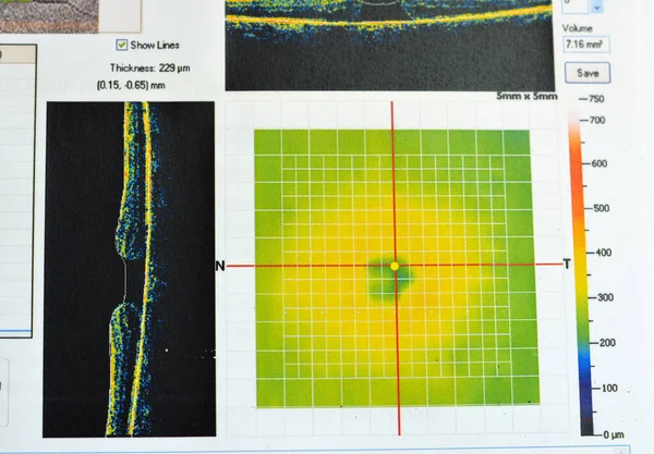
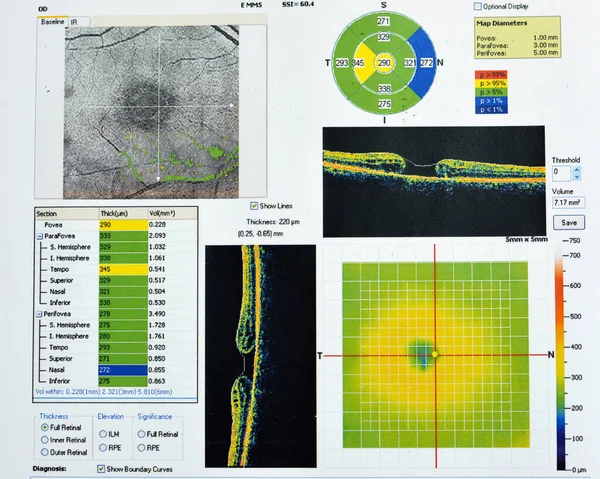
Stock image OCT of the eye reveals faint epimacular membrane and full thickness macular hole involving the fovea, surrounding diffuse macular oedema showing few cystoid changes for follow up, selective focus
Published:
May.17, 2023 13:30:50
Author:
Tamer_Soliman
Views:
6
Downloads:
1
File type:
image
/
jpg
File size:
11.49 MB
Orginal size:
4800
x
3840
px
Available sizes:
S
(500 x 400)
M
(1000 x 800)
L
(2000 x 1600)
XL
(4800 x 3840)
EL0
(4800 x 3840)
Level:
beginner
Download Image
Image topics:
Image
Blood
Medicine
Medical
Care
Eyes
Eye
Hospital
Visual
Pressure
Patient
Optical
Lens
Hole
Globe
Fluid
Vision
Vein
Optic
Sick
Membrane
Accumulation
Holes
Ophthalmic
Ophthalmologist
Ocular
Ophthalmology
Eyeball
Vessels
Cataract
Diabetes
Retina
Nerve
Retinal
Atrophy
Glaucoma
Acuity
Oct
Fovea
Fundus
Macula
Macular
Retinopathy
Edema
Exudate
Oedema
Follow Up
Sequelae
Epimacular
Similar stock images
OCT Of The Eye Reveals Faint Epimacular Membrane And Full Thickness Macular Hole Involving The Fovea, Surrounding Diffuse Macular Oedema Showing Few Cystoid Changes For Follow Up, Selective Focus
5242 × 3847
OCT Of The Eye Reveals Faint Epimacular Membrane And Full Thickness Macular Hole Involving The Fovea, Surrounding Diffuse Macular Oedema Showing Few Cystoid Changes For Follow Up, Selective Focus
5048 × 3895
OCT Of The Eye Reveals Faint Epimacular Membrane And Full Thickness Macular Hole Involving The Fovea, Surrounding Diffuse Macular Oedema Showing Few Cystoid Changes For Follow Up, Selective Focus
5136 × 3738
OCT Of The Eye Reveals Faint Epimacular Membrane And Full Thickness Macular Hole Involving The Fovea, Surrounding Diffuse Macular Oedema Showing Few Cystoid Changes For Follow Up, Selective Focus
5561 × 3708
OCT Of The Eye Reveals Faint Epimacular Membrane And Full Thickness Macular Hole Involving The Fovea, Surrounding Diffuse Macular Oedema Showing Few Cystoid Changes For Follow Up, Selective Focus
5879 × 3536
OCT Of The Eye Reveals Faint Epimacular Membrane And Full Thickness Macular Hole Involving The Fovea, Surrounding Diffuse Macular Oedema Showing Few Cystoid Changes For Follow Up, Selective Focus
5517 × 3641
OCT Of The Eye Reveals Faint Epimacular Membrane And Full Thickness Macular Hole Involving The Fovea, Surrounding Diffuse Macular Oedema Showing Few Cystoid Changes For Follow Up, Selective Focus
5430 × 3774
Optical Coherence Tomography (OCT) Showing The Optic Nerve Of A Patient At Risk For Glaucoma.
3558 × 4217