Home
Images
Vectors
Videos
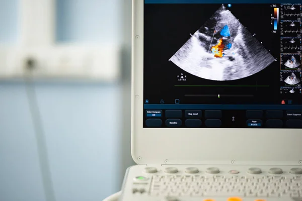
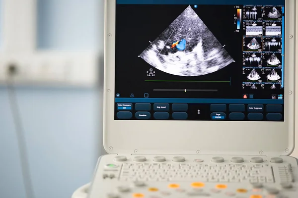
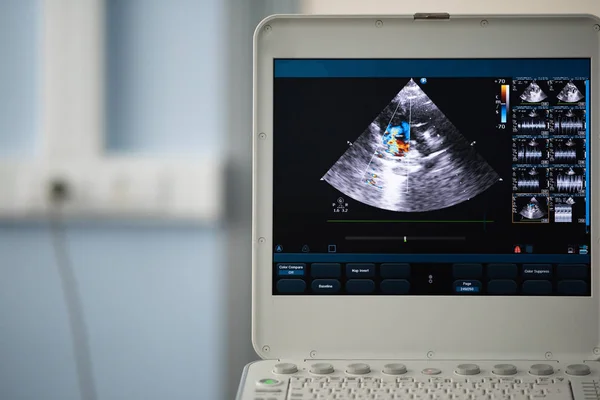
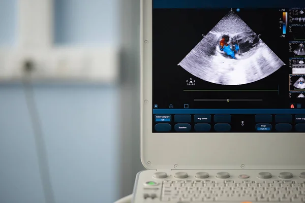
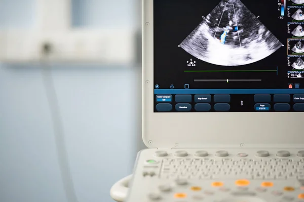
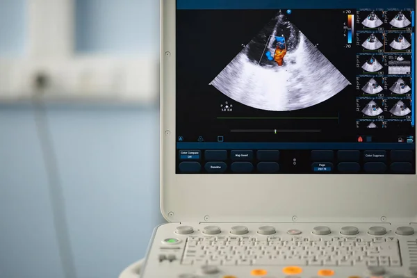
Stock image On the screen of a modern ultrasound scanner, an image of the heart cavities with red and blue streams of blood regurgitation depicted by the Doppler method.
Published:
Jul.23, 2018 09:08:03
Author:
Faustasyan
Views:
7
Downloads:
1
File type:
image
/
jpg
File size:
10.59 MB
Orginal size:
5500
x
3670
px
Available sizes:
S
(500 x 334)
M
(1000 x 667)
L
(2000 x 1335)
XL
(5500 x 3670)
EL0
(5500 x 3670)
Level:
beginner
Download Image
Image topics:
Image
Colors
Computer
Focus
Equipment
Blood
Healthcare
Medical
Care
Procedure
Technology
Instrument
Machine
Heart
Hospital
Device
Screen
Monitor
Visual
Pulse
Tool
Science
Trace
Analyzing
Valve
Vein
Diagnosis
Examination
Imaging
Scan
Diagnostic
Scanner
Scanning
Cardiologist
Cavity
Artery
Ultrasound
Atrium
Ultrasonic
Doppler
Aorta
Ventricle
Sonography
Valvular
Echocardiography
Septum
Mitral
Tricuspid
Stenosis
Echocardiogram
Similar stock images
On The Screen Of The Ultrasound Scanner, The Heart Image In The Four-chamber Position And Marked By The Blue And Red Doppler Method Are Valve Regurgitation.
3500 × 2336
On The Ultrasound Screen, The Image Of The Heart In The Four-chamber Position And The Doppler Method Shows Tricuspid Regurgitation.
3500 × 2336
On The Ultrasound Screen, The Doppler Method Depicts Regurgitation On The Valve.
6000 × 4005
On The Screen Of A Modern Ultrasound Scanner, An Image Of The Heart Cavities With Red And Blue Streams Of Blood Regurgitation Depicted By The Doppler Method.
5500 × 3670
On The Screen Of A Modern Ultrasound Scanner, An Image Of The Heart Cavities With Red And Blue Streams Of Blood Regurgitation Depicted By The Doppler Method.
6000 × 4005
On The Screen Of An Ultrasound Device, A Heart Scan In A Four-chamber Position With A Doppler Image Of Tricuspid Regurgitation.
6000 × 4004
On The Screen Of The Ultrasound Scanner, The Heart Image In The Four-chamber Position And Marked By The Blue And Red Doppler Method Are Valve Regurgitation.
4500 × 3003
On The Screen Of The Ultrasound Scanner, The Heart Image In The Four-chamber Position And Marked By The Blue And Red Doppler Method Are Valve Regurgitation.
5500 × 3671