photo free
Home Images Vectors Videos

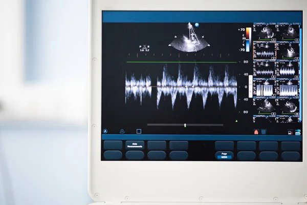
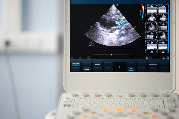
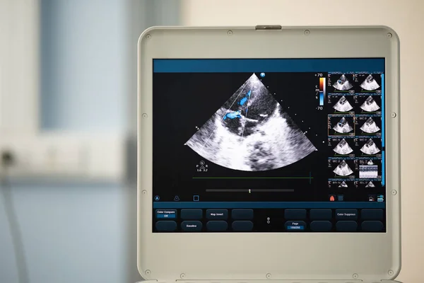
Stock image On the screen of the ultrasound apparatus, the scan of the left ventricle of the heart in the position for measuring the ejection fraction by Teyolz.
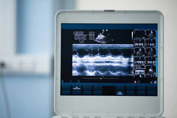
Stock image On the screen of the ultrasound apparatus, the scan of the left ventricle of the heart in the position for measuring the ejection fraction by Teyolz.
Published: Jul.23, 2018 09:02:51
Author: Faustasyan
Views: 7
Downloads: 0
File type: image / jpg
File size: 15.35 MB
Orginal size: 6000 x 4004 px
Available sizes:
S
(500 x 334)
M
(1000 x 667)
L
(2000 x 1335)
XL
(6000 x 4004)
EL0
(6000 x 4004)
Level: beginner
Download Image
Image topics:

Similar stock images

stock image The ultrasound screen with the image of the heart cut, on which the gradient of the right ventricular outflow tract was measured.

The Ultrasound Screen With The Image Of The Heart Cut, On Which The Gradient Of The Right Ventricular Outflow Tract Was Measured.

5000 × 3337
stock image The screen of the ultrasound device with the image of measuring the pressure gradient

The Screen Of The Ultrasound Device With The Image Of Measuring The Pressure Gradient

6000 × 4004
stock image On the screen of the ultrasound scanner, the image of the heart in the four-chamber position and marked by the blue doppler method is mitral regurgitation.

On The Screen Of The Ultrasound Scanner, The Image Of The Heart In The Four-chamber Position And Marked By The Blue Doppler Method Is Mitral Regurgitation.

5000 × 3337
stock image On the screen of the ultrasound machine, the image of the right ventricular outflow tract with the established control volume at the level of the pulmonary valve and the pulse pressure gradient measurement between the cavities by the pulse doppler me

On The Screen Of The Ultrasound Machine, The Image Of The Right Ventricular Outflow Tract With The Established Control Volume At The Level Of The Pulmonary Valve And The Pulse Pressure Gradient Measurement Between The Cavities By The Pulse Doppler Me

5000 × 3337
stock image Screen of an ultrasound device with a heart scan. Using the Doppler method, the right ventricular outflow tract is depicted.

Screen Of An Ultrasound Device With A Heart Scan. Using The Doppler Method, The Right Ventricular Outflow Tract Is Depicted.

6000 × 4004
stock image On the ultrasound screen, the image of the heart in the four-chamber position and the Doppler method shows tricuspid regurgitation.

On The Ultrasound Screen, The Image Of The Heart In The Four-chamber Position And The Doppler Method Shows Tricuspid Regurgitation.

6000 × 4004
stock image On the ultrasound screen, the image of the heart in the four-chamber position and the Doppler method shows tricuspid regurgitation.

On The Ultrasound Screen, The Image Of The Heart In The Four-chamber Position And The Doppler Method Shows Tricuspid Regurgitation.

5000 × 3337
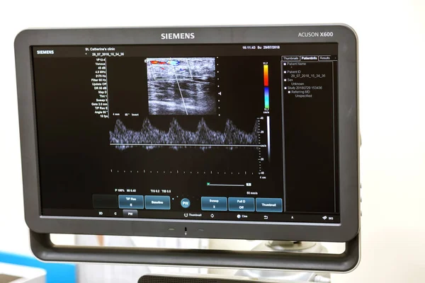
stock image ODESSA, UKRAINE - July 29, 2018: Woman undergoes medical examination of veins on ultrasound sensor in modern technology clinic. Condition of veins and examination of doppler on monitor

ODESSA, UKRAINE - July 29, 2018: Woman Undergoes Medical Examination Of Veins On Ultrasound Sensor In Modern Technology Clinic. Condition Of Veins And Examination Of Doppler On Monitor

5472 × 3648