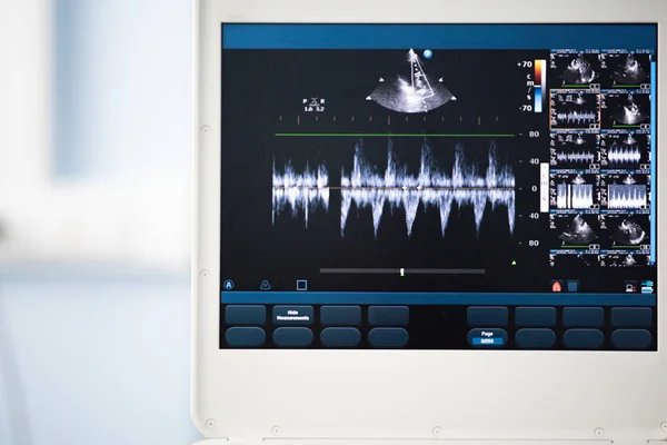
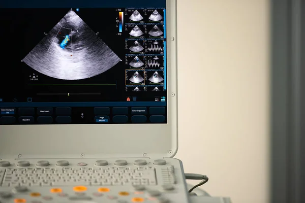
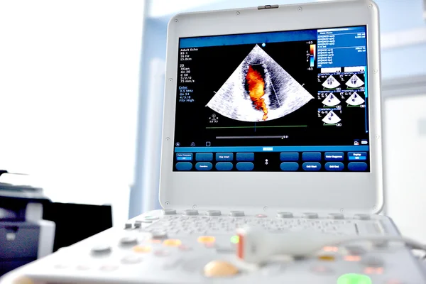
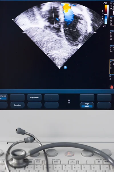
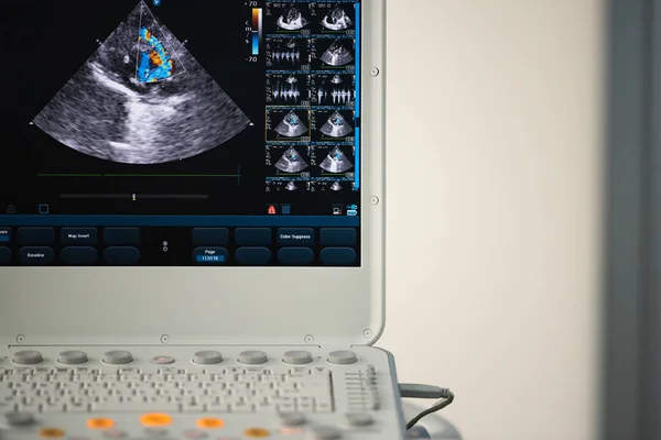
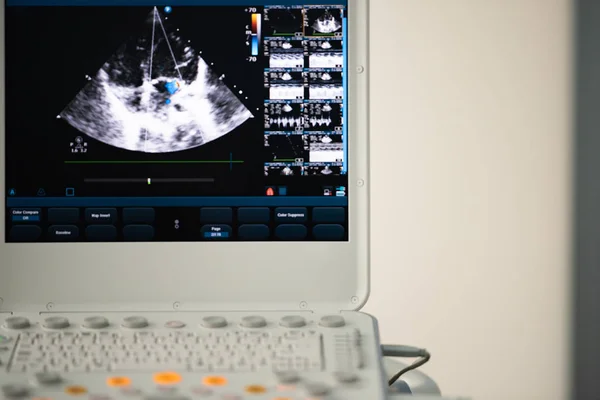
Stock image On the screen of the ultrasound scanner, the image of the heart in the four-chamber position and marked by the blue doppler method is mitral regurgitation.
Published: Jul.23, 2018 09:08:03
Author: Faustasyan
Views: 7
Downloads: 0
File type: image / jpg
File size: 5.4 MB
Orginal size: 4000 x 2669 px
Available sizes:
Level: beginner