Home
Images
Vectors
Videos
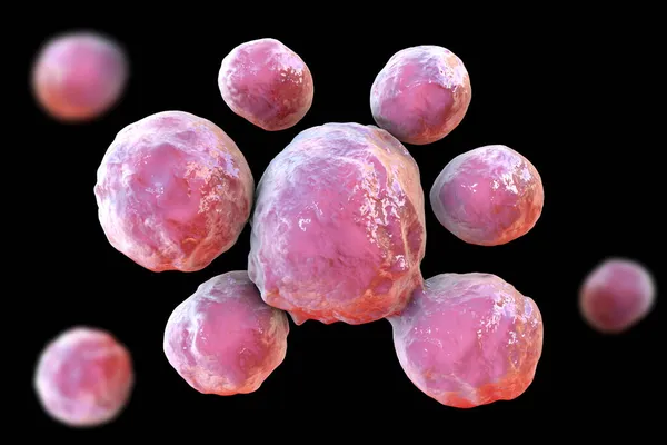
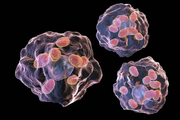
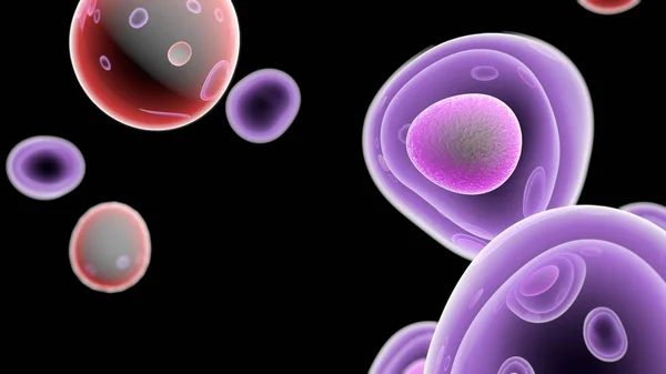
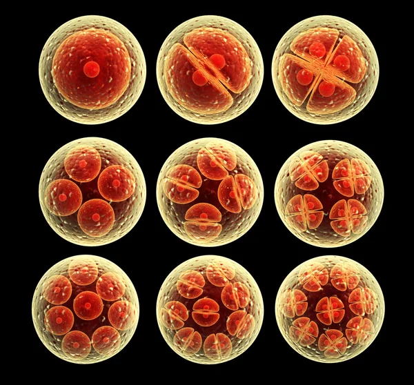
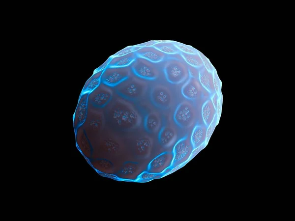
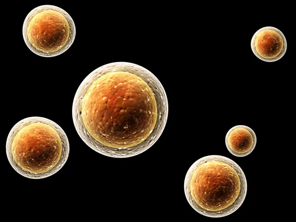
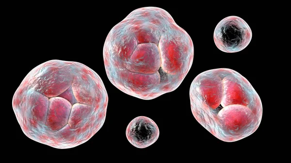
Stock image Prototheca wickerhamii green algae that cause protothecosis, clinically seen as skin nodules and elbow bursitis. 3D illustration shows sporangia with endospores and nonviable ghostlike forms.
Published:
Jan.27, 2023 10:00:08
Author:
katerynakon
Views:
2
Downloads:
0
File type:
image
/
jpg
File size:
6.24 MB
Orginal size:
7200
x
4050
px
Available sizes:
S
(500 x 281)
M
(1000 x 563)
L
(2000 x 1125)
XL
(7200 x 4050)
EL0
(7200 x 4050)
Level:
silver
Download Image
Image topics:
Illustration
Isolated
Shape
Human
Environment
Medicine
Medical
3d
Skin
Biological
Education
Biology
Science
Tissue
Elbow
Research
Joint
Magnification
Scientific
Dermatology
Neglected
Diagnosis
Rare
Spore
Microscope
Smear
Algae
Infection
Diagnostics
Subcutaneous
Microscopy
Bursa
Cutaneous
Black Background
Sporangium
Sporangia
Bursitis
Hyperkeratosis
Parakeratosis
Acanthosis
Endospores
Prototheca Wickerhamii
Prototheca
Protothecosis
Achlorophyllous
Systemic Infection
Similar stock images
Bacteria Coxiella Burnetii, 3D Illustration. Gram-negative Bacteria That Cause Q Fever, Are Transmitted To Humans From Sheep, Goats And Cattle
6000 × 4000
Fungi Paracoccidioides Lutzii, A Dimorphic Fungus That Causes Paracoccidioidomycosis Found In Brazil And Ecuador. Scientific 3D Illustration Showing Characteristic Morphology Of The Fungus
6000 × 4000
Macrophages Infected By Leishmania Amastigotes
4500 × 3000
Plague Bacterium Yersinia Pestis, Scientifically Accurate 3D Illustration Showing Structure Of The Cell With DNA, Plasmids And Ribosomes
5635 × 3757
T Cells Attacking Cancer Cells- 3D Illustration
3840 × 2160
Process Division Of Cell
4024 × 3750
3d Rendering - Nematode Worm Egg
4836 × 3628
Colony Of Viruses
5000 × 3750