Home
Images
Vectors
Videos
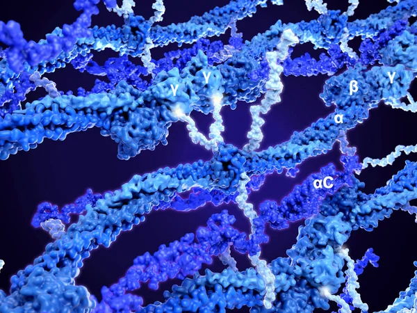
Stock image Structure of a fibrin polymer. A fibrin monomer (highlighted) is composed of 2, 2, 2 and 2C domains. There are 4 flexible chains (light blue) in the center. The 2 short .chains bind to two adjacent domains of the polymer.
Published:
Jun.01, 2020 10:42:48
Author:
animaxx3d
Views:
108
Downloads:
3
File type:
image
/
jpg
File size:
8.41 MB
Orginal size:
8000
x
6000
px
Available sizes:
S
(500 x 375)
M
(1000 x 750)
L
(2000 x 1500)
XL
(8000 x 6000)
EL0
(8000 x 6000)
Level:
bronze
Download Image
Image topics:
Protein
Blood
Clot
Stroke
Molecule
Polymer
Hemorrhage
Platelet
Fibrin
Thrombus
Thrombosis
Hemophilia
Polymerization
Hemostasis
Thrombin
Blood Clotting
Fibrinogen
Coagulation Cascade
Similar stock images
Infection Virus Bacteria Cell Micro Microscope Inside Interior Human Body Section Movement Reaction. Cell Aggregation And Gene Mutations. Medical Science Experiments In The Lab. 3D Illustration.
8000 × 5336
DNA Research Background
4300 × 2580
Helix Of DNA
8000 × 4500
Helix Of DNA
8000 × 5000
DNA Research Background
4300 × 2580
Ribonucleic Acid Strands Consisting Of Nucleotides Important For Protein Bio-synthesis - 3d Illustration
8500 × 4250
Biotechnology Genetic Research
4300 × 3010