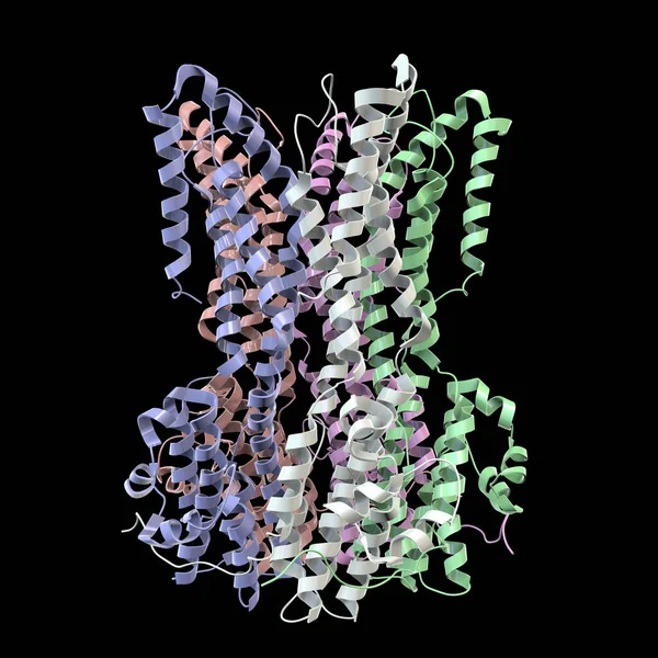
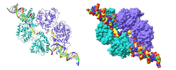
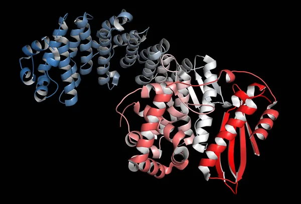
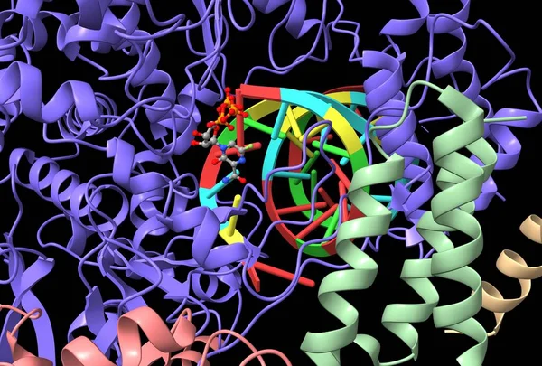
Stock image Structure of Favipiravir bound to replicating polymerase complex of SARS-CoV-2 in the pre-catalytic state, 3D cartoon model with active center close-up, based on PDB 7ctt, black background
Published: Jun.30, 2021 11:54:13
Author: unnaugan
Views: 7
Downloads: 0
File type: image / jpg
File size: 2.94 MB
Orginal size: 6000 x 4056 px
Available sizes:
Level: beginner