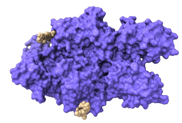
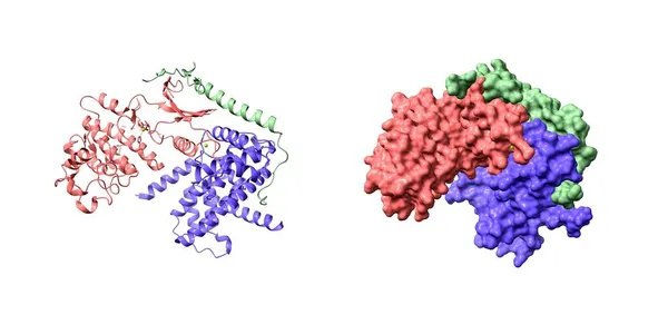
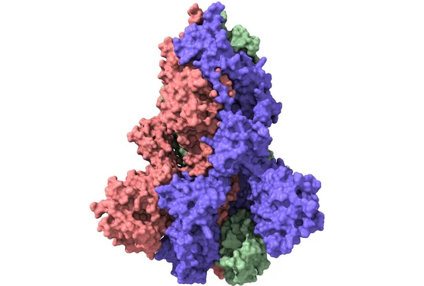
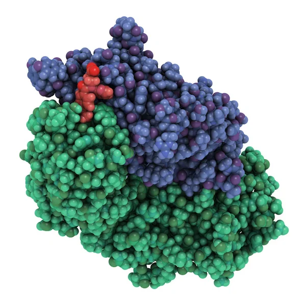
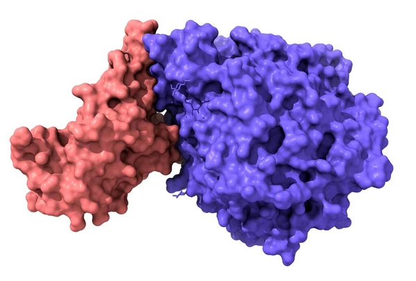
Stock image Structure of novel coronavirus spike receptor-binding domain (pink) complexed with its receptor ACE2 (blue), white background
Published: Jun.23, 2020 06:42:42
Author: unnaugan
Views: 5
Downloads: 1
File type: image / jpg
File size: 0.87 MB
Orginal size: 5000 x 3380 px
Available sizes:
Level: beginner