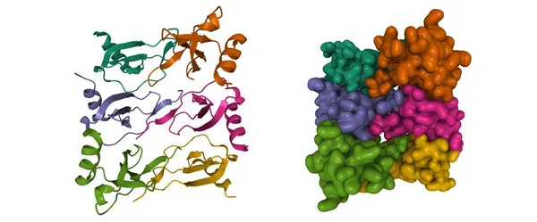
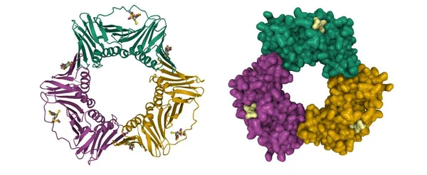
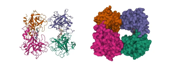
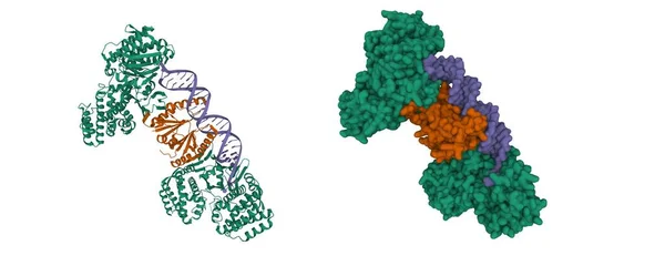
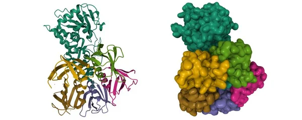
Stock image Structure of shiga toxin, B-chain pentamer and A-chain subunit (green), 3D cartoon and Gaussian surface model, white background
Published: Jun.14, 2021 13:04:23
Author: unnaugan
Views: 2
Downloads: 0
File type: image / jpg
File size: 5.27 MB
Orginal size: 10000 x 4000 px
Available sizes:
Level: beginner