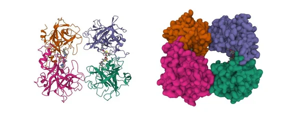
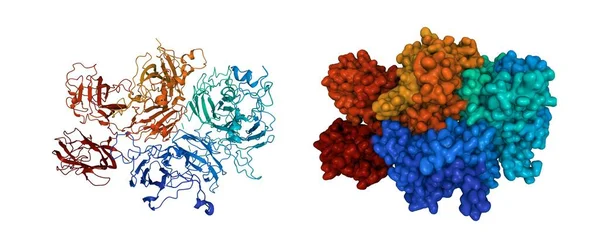
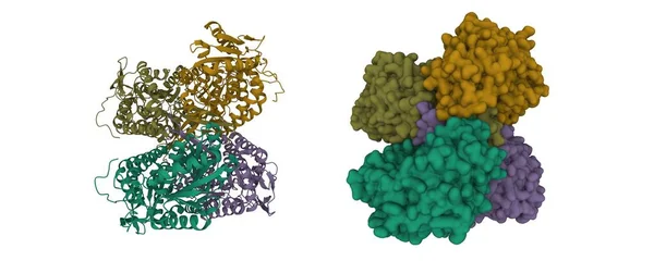
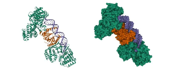
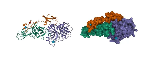
Stock image Structure of tissue factor (green) -factor VIIa (brown and violet) complex, 3D cartoon and Gaussian surface models, chain id color scheme, based on PDB 1j9c, white background
Published: Jul.29, 2021 08:53:26
Author: unnaugan
Views: 21
Downloads: 1
File type: image / jpg
File size: 3.57 MB
Orginal size: 10000 x 4100 px
Available sizes:
Level: beginner