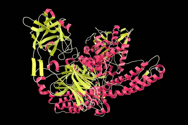
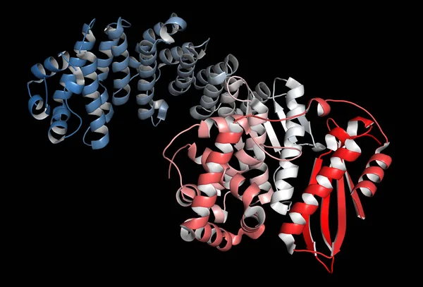
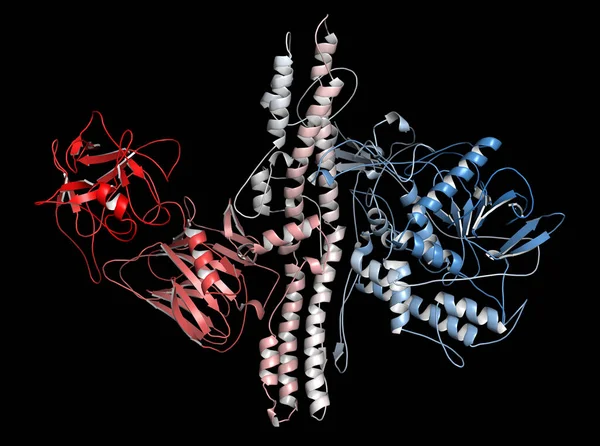
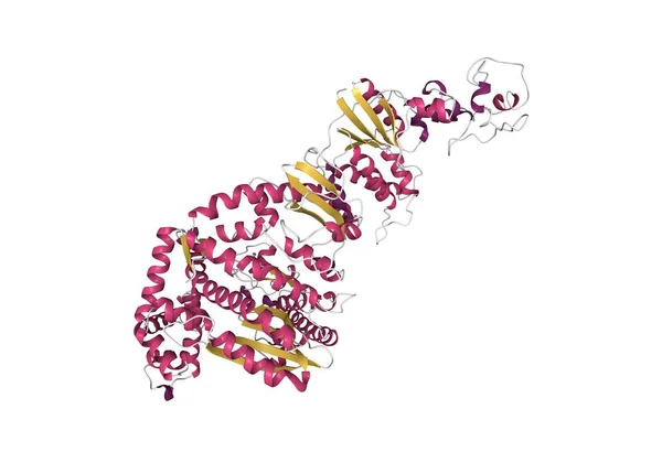
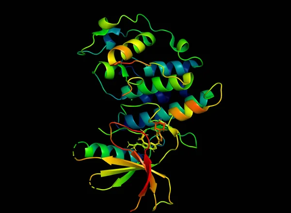
Stock image Tertiary structure of human vitamin D binding protein with the differently colored secondary structure elements, 3D cartoon model, black background
Published: Oct.05, 2020 07:00:06
Author: unnaugan
Views: 4
Downloads: 0
File type: image / jpg
File size: 2.81 MB
Orginal size: 4096 x 4096 px
Available sizes:
Level: beginner