Home
Images
Vectors
Videos
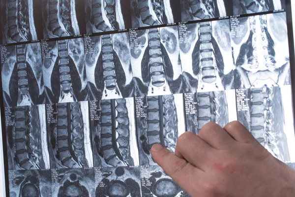
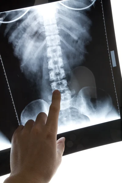
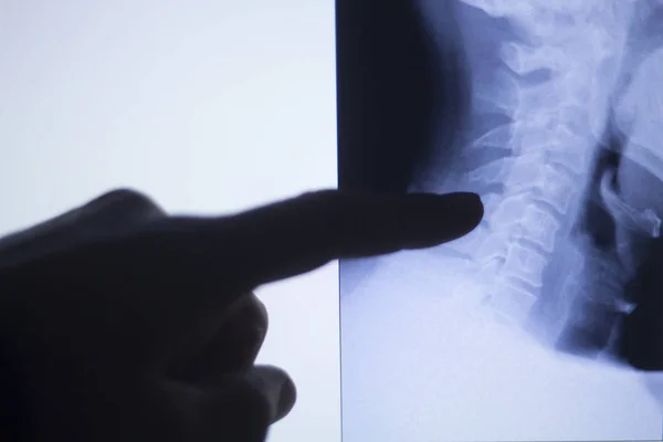
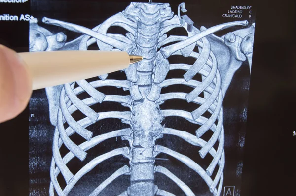
Stock image Thoracic spine MRI or CT X-ray image. The doctor points to the thoracic spine X-ray image of the patient, where it is often localized to the thoracic spine osteochondrosis and other diseases
Published:
Sep.27, 2017 06:33:49
Author:
Shidlovski
Views:
824
Downloads:
7
File type:
image
/
jpg
File size:
9.68 MB
Orginal size:
6016
x
4000
px
Available sizes:
S
(500 x 332)
M
(1000 x 665)
L
(2000 x 1330)
XL
(6016 x 4000)
EL0
(6016 x 4000)
Level:
bronze
Download Image
Image topics:
Image
Scene
Illness
Medical
Cord
Back
Concept
Idea
Doctor
Bone
Patient
Neck
Disease
Cancer
Film
Point
Pain
Column
Spine
Magnetic
Skeleton
Diagnosis
Anatomy
Spinal
X Ray
Surgery
Injury
Scan
Diagnostic
Disorder
Compress
Cavity
Radiology
Xray
Upper
Inflammation
Mri
Thorax
Thoracic
Ct
Neurosurgery
Cervical
Vertebral
Neuroscience
Vertebrae
Osteochondrosis
Neuropathy
Localized
Metastasis
Myelopathy
Similar stock images
Sternum And Thymus Gland. Doctor Points Out Ballpoint Pen On Sternum And Location Of Thymus Gland As Organ Of Immune And Lymphatic Systems On Child 3D Visualization Of CT Or MRI Diagnosis Scans
6016 × 4000
Doctor Hand Shows Something On MRI Results (magnetic Resonance T
6000 × 4000
Doctor Examines X-ray Picture Of Human Spine And Chest
6240 × 4160
Doctor Examines X-ray Picture Of Human Spine
6240 × 4160
Doctor Examines X-ray Picture Of Human Spine
6240 × 4160
X-ray Image
2912 × 4368
Back Spine Vertabrae Xray Scan
5184 × 3456
Doctor Hand Shows Something On The MRI Results Magnetic Resonance Therapy
5500 × 3529