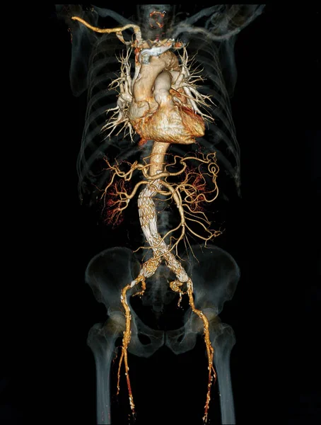
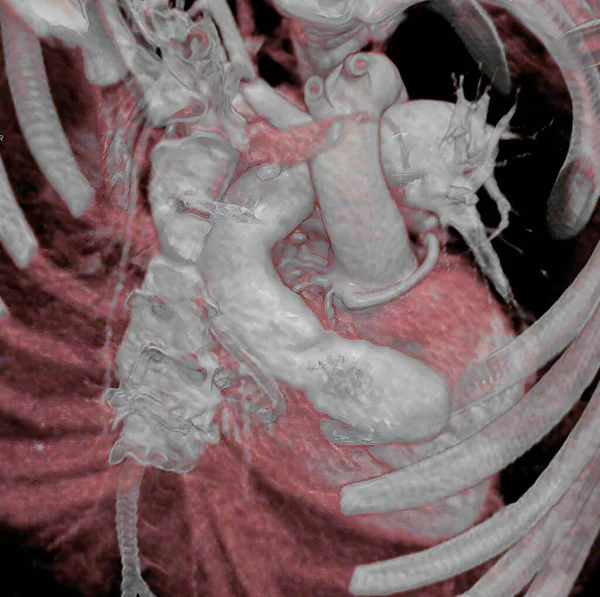
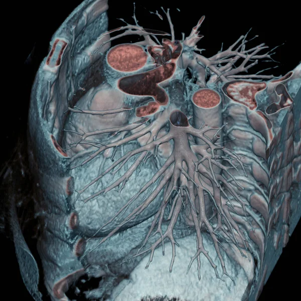
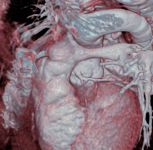
Stock image Thorax: 3D CT scan.

Published: Aug.16, 2022 14:04:07
Author: imagepointfr
Views: 6
Downloads: 0
File type: image / jpg
File size: 12.53 MB
Orginal size: 4984 x 5015 px
Available sizes:
Level: silver