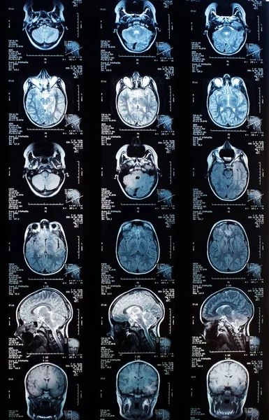
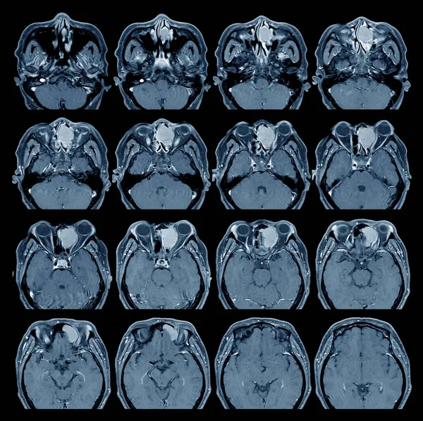
Stock image Tomography of the brain obtained by scanner.

Published: May.03, 2024 04:29:01
Author: imagepointfr
Views: 0
Downloads: 0
File type: image / jpg
File size: 21.64 MB
Orginal size: 4101 x 5440 px
Available sizes:
Level: silver