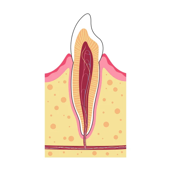
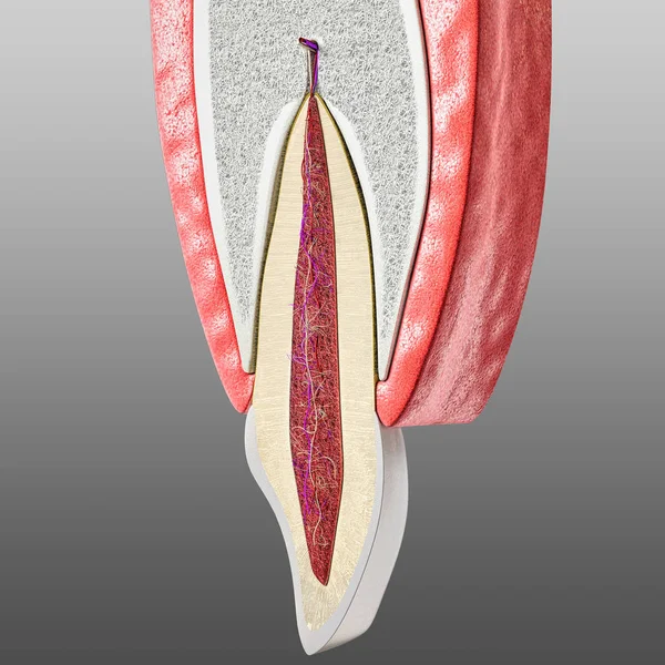
Stock image tooth and periodontium anatomy. Sectional human central incisor showing the anatomical structures that form the dental tissue and the periodontal tissues. Infographic, 3D illustration
Published: May.14, 2019 08:10:53
Author: p.turrini
Views: 183
Downloads: 2
File type: image / jpg
File size: 8.39 MB
Orginal size: 4000 x 4000 px
Available sizes:
Level: beginner