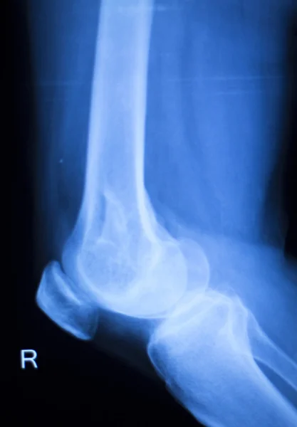
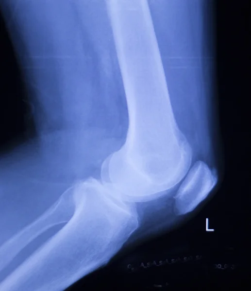
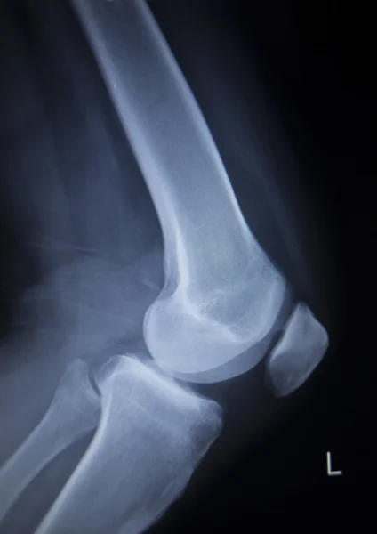
Stock image X-ray Knee join Showing large osteolytic lesuion of medial aspect of left distal femur.with soft tissure mass.and malignant bone tumor,osteosarcoma is suspected.

Published: Oct.11, 2018 08:08:32
Author: Richmanphoto
Views: 173
Downloads: 3
File type: image / jpg
File size: 2.9 MB
Orginal size: 3500 x 4500 px
Available sizes:
Level: bronze